Your Chronic superior mesenteric vein thrombosis ct images are available. Chronic superior mesenteric vein thrombosis ct are a topic that is being searched for and liked by netizens today. You can Find and Download the Chronic superior mesenteric vein thrombosis ct files here. Get all royalty-free vectors.
If you’re searching for chronic superior mesenteric vein thrombosis ct pictures information related to the chronic superior mesenteric vein thrombosis ct topic, you have visit the ideal site. Our website always gives you hints for seeing the highest quality video and picture content, please kindly search and locate more informative video content and images that fit your interests.
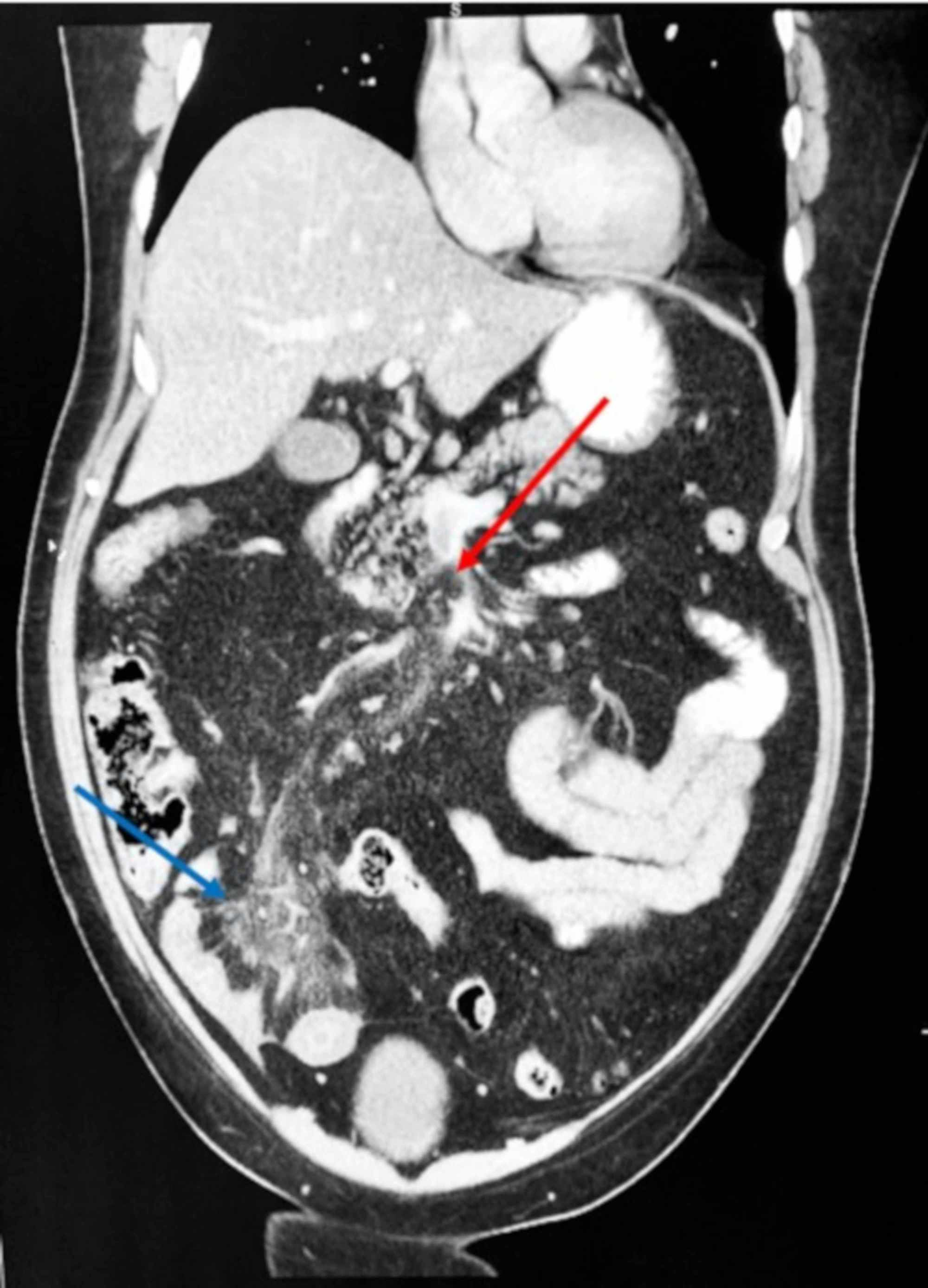
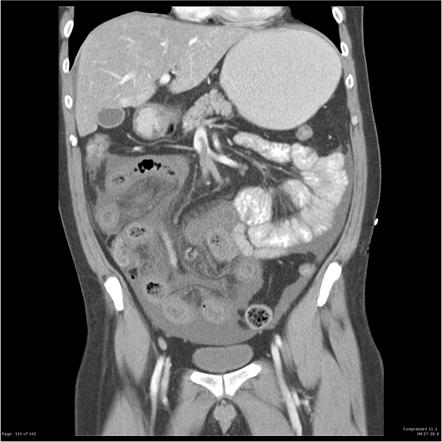
Chronic Superior Mesenteric Vein Thrombosis Ct. SAGAR Radiology Department City Hospital NHS Trust Birmingham UK Thrombosis in Acute We describe the computed tomography CT appearances of four patients with acute or acute on chronic case 3 pancreatitis which demonstrated isolated superior mesenteric vein SMV thrombosis. Chronic mesenteric ischemia also known as intestinal angina is most often due to arterial atherosclerotic disease. Chronic mesenteric venous thrombosis accounts for approximately 20 to 40 of total mesenteric venous thrombosis cases and rarely causes intestinal infarction. There is an extension of the thrombus to the portal vein there is a partial filling defect in the anterior wall witch can be seen partially extending to the right branch of the.
 Acute Superior Mesenteric Vein Thrombosis In The Setting Of Cytomegalovirus Mononucleosis A Case Report And Review Of The Literature The Lancet Infectious Diseases From thelancet.com
In three of the four cases follow-up CT scans showed the SMV thrombosis to have. Contrast-enhanced CT diagnoses about 90 of cases. Reversible superior mesenteric vein thrombosis in acute pancreatitis. The most common symptom related to mesenteric venous thrombosis was abdominal pain but vomiting diarrhea and fever were also considered possible related symptoms. CT ABDOMEN W CON—–554000 ULTRASOUND 15 MIN—–21000. Associated portal venous thrombosis can be seen if the disease originates in the major.
The portal vein is formed by the confluence of the splenic and superior mesenteric veins which drain the spleen and small intestine respectively figure 1.
The vein has an internal hypodensity in the portalvenous phase. Chronic mesenteric venous thrombosis accounts for approximately 20 to 40 of total mesenteric venous thrombosis cases and rarely causes intestinal infarction. Occlusion of the portal vein by thrombus portal vein thrombosis PVT typically occurs in patients with cirrhosis andor prothrombotic disorders table 1. The diagnosis of mesenteric vein thrombosis relies heavily on imaging. Furthermore bowel ischemia may not develop immediately. Treatment could include antibiotics anticoagulants surgery to remove the clot or to place drugs to dissolve the clot or a small intestine resection.
 Source: researchgate.net
Occlusion of the portal vein by thrombus portal vein thrombosis PVT typically occurs in patients with cirrhosis andor prothrombotic disorders table 1. Mesenteric vein thrombosis almost always involves the distal small intestine superior mesenteric venous drainage and rarely involves the colon inferior mesenteric venous drainage. Symptoms when the diagnosis of acute mesenteric venous thrombosis was established as well as symptoms at the chronic stage if any were reported. Its branches are patent. A thrombosis of the superior mesenteric vein SMV.
 Source: cmaj.ca
He had contracted COVID-19 9 days prior. Further investigation ruled out haematological causes and COVID-19 was. SAGAR Radiology Department City Hospital NHS Trust Birmingham UK Thrombosis in Acute We describe the computed tomography CT appearances of four patients with acute or acute on chronic case 3 pancreatitis which demonstrated isolated superior mesenteric vein SMV thrombosis. The portal vein is formed by the confluence of the splenic and superior mesenteric veins which drain the spleen and small intestine respectively figure 1. The superior mesenteric vein is often involved whereas involvement of the inferior mesenteric vein is rare.
 Source: thelancet.com
The most common symptom related to mesenteric venous thrombosis was abdominal pain but vomiting diarrhea and fever were also considered possible related symptoms. The inferior mesenteric vein is involved only. CT chest abdomen and pelvis revealed an extensive thrombus extending from the portal vein to the superior mesenteric vein. Filling defect in the portal vein. Occlusion of the portal vein by thrombus portal vein thrombosis PVT typically occurs in patients with cirrhosis andor prothrombotic disorders table 1.

Mesenteric Vein Thrombosis Diagnosis. Filling defect in the portal vein. Mesenteric Vein Thrombosis Diagnosis. The anatomic site of involvement in acute mesenteric venous thrombosis is most often ileum 64 to 83 percent or jejunum 50 to 81 percent followed by colon. When you have mesenteric venous thrombosis MVT you have a blood clot in a vein around where your intestines attach to your belly.
 Source: ejves.com
He had contracted COVID-19 9 days prior. Main portal vein is dilated 145mm. The most practical imaging modality is CT as seen in the image above. The symptoms are usually not typical enough to offer a clinical diagnosis. The superior mesenteric vein is often involved whereas involvement of the inferior mesenteric vein is rare.
 Source: researchgate.net
Associated portal venous thrombosis can be seen if the disease originates in the major. Filling defect in the superior mesenteric vein. CT ABDOMEN W CON—–554000 ULTRASOUND 15 MIN—–21000. Chronic mesenteric venous thrombosis accounts for approximately 20 to 40 of total mesenteric venous thrombosis cases and rarely causes intestinal infarction. Reversible superior mesenteric vein thrombosis in acute pancreatitis.
 Source: researchgate.net
Each of the aforementioned conditions re-quires a different approach to diagnosis and management. The symptoms are usually not typical enough to offer a clinical diagnosis. Treatment could include antibiotics anticoagulants surgery to remove the clot or to place drugs to dissolve the clot or a small intestine resection. Up to 10 cash back The main aim of this study was to evaluate the association of computed tomography CT findings at admission and bowel resection rate in patients with mesenteric venous thrombosis MVT. Further investigation ruled out haematological causes and COVID-19 was.
 Source: cureus.com
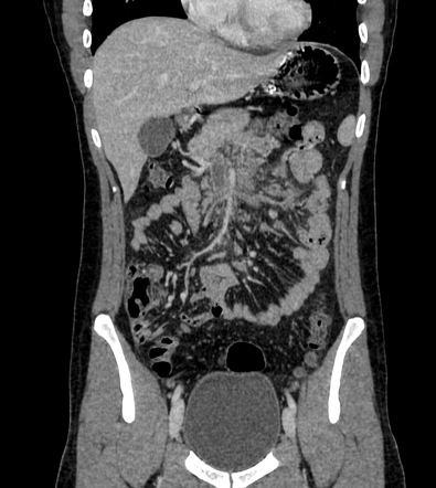
The vein has an internal hypodensity in the portalvenous phase. Chronic mesenteric venous thrombosis is differentiated from acute mesenteric venous thrombosis by the existence of collateral venous circulation and cavernoma around the thrombosed vein. The inferior mesenteric vein is involved only. Mesenteric Vein Thrombosis Diagnosis. This condition is most often diagnosed with a CT scan.
 Source: radiopaedia.org
Acute thrombosis commonly presents with abdominal pain and chronic type with features of portal hypertension. Furthermore bowel ischemia may not develop immediately. Chronic mesenteric venous thrombosis is differentiated from acute mesenteric venous thrombosis by the existence of collateral venous circulation and cavernoma around the thrombosed vein. SAGAR Radiology Department City Hospital NHS Trust Birmingham UK Thrombosis in Acute We describe the computed tomography CT appearances of four patients with acute or acute on chronic case 3 pancreatitis which demonstrated isolated superior mesenteric vein SMV thrombosis. There is an extension of the thrombus to the portal vein there is a partial filling defect in the anterior wall witch can be seen partially extending to the right branch of the.
 Source: radiopaedia.org
The portal vein is formed by the confluence of the splenic and superior mesenteric veins which drain the spleen and small intestine respectively figure 1. Symptoms when the diagnosis of acute mesenteric venous thrombosis was established as well as symptoms at the chronic stage if any were reported. The superior mesenteric vein is often involved whereas involvement of the inferior mesenteric vein is rare. The presence of collateral circulation and cavernoma around a chronically thrombosed vein differentiates chronic from acute disease. Each of the aforementioned conditions re-quires a different approach to diagnosis and management.
 Source: thoracickey.com
The portal vein is formed by the confluence of the splenic and superior mesenteric veins which drain the spleen and small intestine respectively figure 1. Filling defect in the portal vein. The superior mesenteric vein is often involved whereas involvement of the inferior mesenteric vein is rare. Filling defect in the superior mesenteric vein. Chronic mesenteric ischemia also known as intestinal angina is most often due to arterial atherosclerotic disease.
 Source: radiopaedia.org
Contrast-enhanced CT diagnoses about 90 of cases. Mesenteric vein thrombosis is increasingly recognized as a cause of mesenteric ischemia. Furthermore bowel ischemia may not develop immediately. Symptoms when the diagnosis of acute mesenteric venous thrombosis was established as well as symptoms at the chronic stage if any were reported. The inferior mesenteric vein is involved only.
 Source: radiopaedia.org
Mesenteric vein thrombosis is increasingly recognized as a cause of mesenteric ischemia. Treatment could include antibiotics anticoagulants surgery to remove the clot or to place drugs to dissolve the clot or a small intestine resection. Symptoms when the diagnosis of acute mesenteric venous thrombosis was established as well as symptoms at the chronic stage if any were reported. Reversible superior mesenteric vein thrombosis in acute pancreatitis. There is an extension of the thrombus to the portal vein there is a partial filling defect in the anterior wall witch can be seen partially extending to the right branch of the.
 Source: radiopaedia.org
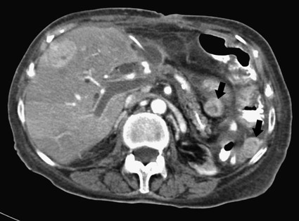
The most practical imaging modality is CT as seen in the image above. McGovern Medical School Portal and Splenic Vein Angioplasty. Chronic mesenteric venous thrombosis is differentiated from acute mesenteric venous thrombosis by the existence of collateral venous circulation and cavernoma around the thrombosed vein. Filling defect in the portal vein. A 68-year-old man was referred to the general surgeons on account of his abdominal pain of unknown cause.
 Source: radiologykey.com
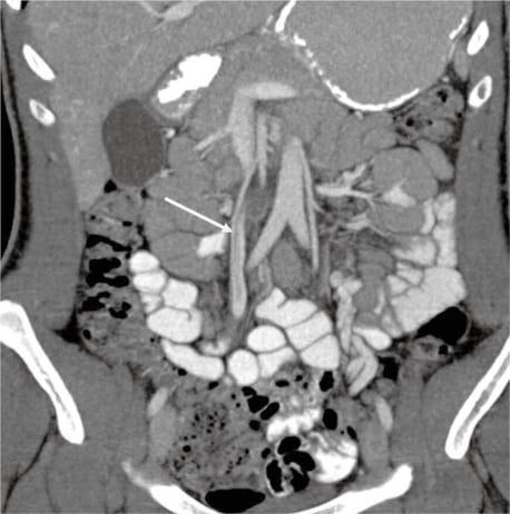
Symptoms when the diagnosis of acute mesenteric venous thrombosis was established as well as symptoms at the chronic stage if any were reported. McGovern Medical School Portal and Splenic Vein Angioplasty. Associated portal venous thrombosis can be seen if the disease originates in the major. A thrombosis of the superior mesenteric vein SMV. The most practical imaging modality is CT as seen in the image above.
 Source: angiologist.com
We describe the computed tomography CT appearances of four patients with acute or acute on chronic case 3 pancreatitis which demonstrated isolated superior mesenteric vein SMV thrombosis. This thrombus has a density of 35 -40 HU. The inferior mesenteric vein is involved only. Chronic mesenteric ischemia also known as intestinal angina is most often due to arterial atherosclerotic disease. Up to 10 cash back The main aim of this study was to evaluate the association of computed tomography CT findings at admission and bowel resection rate in patients with mesenteric venous thrombosis MVT.
 Source: westjem.com
The anatomic site of involvement in acute mesenteric venous thrombosis is most often ileum 64 to 83 percent or jejunum 50 to 81 percent followed by colon. Mesenteric Vein Thrombosis Diagnosis. When you have mesenteric venous thrombosis MVT you have a blood clot in a vein around where your intestines attach to your belly. Symptoms can include fever nausea blood in the stool abdominal distention or pain and vomiting blood. The splenic vein is dilated 103mm.
 Source: radiopaedia.org
The CT Appearances P. The most practical imaging modality is CT as seen in the image above. McGovern Medical School Portal and Splenic Vein Angioplasty. The vein has an internal hypodensity in the portalvenous phase. This is a direct sign of thrombosis.
This site is an open community for users to submit their favorite wallpapers on the internet, all images or pictures in this website are for personal wallpaper use only, it is stricly prohibited to use this wallpaper for commercial purposes, if you are the author and find this image is shared without your permission, please kindly raise a DMCA report to Us.
If you find this site helpful, please support us by sharing this posts to your preference social media accounts like Facebook, Instagram and so on or you can also bookmark this blog page with the title chronic superior mesenteric vein thrombosis ct by using Ctrl + D for devices a laptop with a Windows operating system or Command + D for laptops with an Apple operating system. If you use a smartphone, you can also use the drawer menu of the browser you are using. Whether it’s a Windows, Mac, iOS or Android operating system, you will still be able to bookmark this website.


