Your Chronic superior mesenteric vein thrombosis radiology images are ready in this website. Chronic superior mesenteric vein thrombosis radiology are a topic that is being searched for and liked by netizens today. You can Get the Chronic superior mesenteric vein thrombosis radiology files here. Download all free vectors.
If you’re searching for chronic superior mesenteric vein thrombosis radiology pictures information connected with to the chronic superior mesenteric vein thrombosis radiology topic, you have visit the right site. Our site always gives you suggestions for seeing the highest quality video and picture content, please kindly surf and locate more enlightening video content and graphics that match your interests.
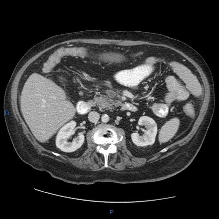
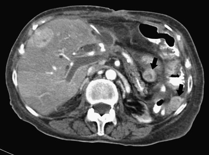
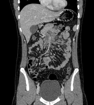
Chronic Superior Mesenteric Vein Thrombosis Radiology. Chronic mesenteric venous thrombosis is differentiated from acute mesenteric venous thrombosis by the existence of collateral venous circulation and cavernoma around the thrombosed vein. Furthermore bowel ischemia may not develop immediately. Branching of mesenteric vein 1 2 can be seen in a and c. MVT accounts for 1 in 5000 to 15000 inpa - tient admissions and 1 in 1000 emer-.
 Superior Mesenteric Venous Thrombosis Radiology Reference Article Radiopaedia Org From radiopaedia.org
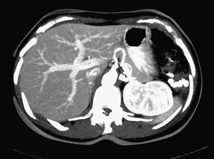
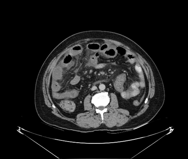
CT chest abdomen and pelvis revealed an extensive thrombus extending from the portal vein to the superior mesenteric vein. The acute mesenteric venous thrombosis MDCT signlow-attenuated intraluminal filling defect at the venous phaseis widely described and accepted in the literature 9 10 but there are few studies presenting chronic radiologic signs of mesenteric venous thrombosis 9 11 12 and there does not appear to be any study evaluating the evolution of the radiologic aspects of acute. The most practical imaging. A Contrast-enhanced maximum intensity projection magnetic resonance MR angiogram reveals collateral vessels from a superior mesenteric vein branch via submucosal varices into liver. When you have mesenteric venous thrombosis MVT you have a blood clot in a vein around where your intestines attach to your belly. Mesenteric vein thrombosis almost always involves the distal small intestine superior mesenteric venous drainage and rarely involves the colon inferior mesenteric venous drainage.
10 Further recurrent bleeding was.
Chronic mesenteric venous thrombosis patients are often asymptomatic with a diagnosis of mesenteric venous thrombosis resulting from incidental findings or portal hypertension. 12 Clinically there are two subtypes of mesenteric ischemia. Chronic mesenteric venous thrombosis accounts for approximately 20 to 40 of total mesenteric venous thrombosis cases and rarely causes intestinal infarction. Mesenteric venous arcades which accompany the arteries unite to form the jejunal and ileal veins in the small bowel mesentery and are joined by the tributaries listed below. 10 Further recurrent bleeding was. He had contracted COVID-19 9 days prior.
 Source: radiologykey.com
The diagnosis of mesenteric vein thrombosis relies heavily on imaging. In one retrospective study on 60 patients with chronic thrombosis of PVs or superior mesenteric veins 39 with variceal bleeding 18 with thrombophilia 9 with variceal bleeding received anticoagulation with recanalization of veins in 3 patients whereas none of the patients who were not anticoagulated recanalized the veins. Chronic mesenteric venous thrombosis is differentiated from acute mesenteric venous thrombosis by the existence of collateral venous circulation and cavernoma around the thrombosed vein. Non opacification of main trunk of superior mesenteric vein up to the portosplenic confluence with perivascular fat stranding. A 68-year-old man was referred to the general surgeons on account of his abdominal pain of unknown cause.
 Source: radiopaedia.org
In patients with extension of thrombosis into the superior mesenteric vein and splenic vein andor presence of hypercoagulability decreased VLS measurements were observed compared with historical control subjects. Mesenteric venous thrombosis MVT describes acute subacute or chronic thrombosis of the superior or inferior mesenteric vein or branches. CT chest abdomen and pelvis revealed an extensive thrombus extending from the portal vein to the superior mesenteric vein. In one retrospective study on 60 patients with chronic thrombosis of PVs or superior mesenteric veins 39 with variceal bleeding 18 with thrombophilia 9 with variceal bleeding received anticoagulation with recanalization of veins in 3 patients whereas none of the patients who were not anticoagulated recanalized the veins. In patients with chronic PVT GI ischemia is frequent.
 Source: radiopaedia.org
Contrast enhanced CT scan of abdomen is quite accurate for diagnosing and differentiating two types of mesenteric venous thrombosis. Contrast enhanced CT scan of abdomen is quite accurate for diagnosing and differentiating two types of mesenteric venous thrombosis. 1 2 Extension to mesenteric venous arches causes intestinal infarction with a reported mortality of up to 50. When you have mesenteric venous thrombosis MVT you have a blood clot in a vein around where your intestines attach to your belly. Its branches are patent.
 Source: researchgate.net
Mesenteric venous thrombosis MVT describes acute subacute or chronic thrombosis of the superior or inferior mesenteric vein or branches. Chronic mesenteric venous thrombosis accounts for approximately 20 to 40 of total mesenteric venous thrombosis cases and rarely causes intestinal infarction. Four patients recovered without radiologic sequelae and 16 developed chronic mesenteric venous thrombosis signs. The most practical imaging. VLS enables objective and quantitative determination of GI mucosal ischemia.
 Source: radiologykey.com
Mesenteric venous arcades which accompany the arteries unite to form the jejunal and ileal veins in the small bowel mesentery and are joined by the tributaries listed below. 3 4 Without recanalization a cavernoma develops associated with a permanent risk of potentially fatal. 2 The modality provides. Venous causes of acute mesenteric ischemia are less common 515 of cases 49 and are most often the result of a thrombosis of the superior mesenteric vein SMV. The symptoms are usually not typical enough to offer a clinical diagnosis.
 Source: cureus.com
A 68-year-old man was referred to the general surgeons on account of his abdominal pain of unknown cause. Four patients recovered without radiologic sequelae and 16 developed chronic mesenteric venous thrombosis signs. Chronic mesenteric venous thrombosis accounts for approximately 20 to 40 of total mesenteric venous thrombosis cases and rarely causes intestinal infarction. Mesenteric vein thrombosis almost always involves the distal small intestine superior mesenteric venous drainage and rarely involves the colon inferior mesenteric venous drainage. Anticoagulation did not influence recovery p 1.
 Source: radiopaedia.org
In patients with extension of thrombosis into the superior mesenteric vein and splenic vein andor presence of hypercoagulability decreased VLS measurements were observed compared with historical control subjects. VLS enables objective and quantitative determination of GI mucosal ischemia. The acute mesenteric venous thrombosis MDCT signlow-attenuated intraluminal filling defect at the venous phaseis widely described and accepted in the literature 9 10 but there are few studies presenting chronic radiologic signs of mesenteric venous thrombosis 9 11 12 and there does not appear to be any study evaluating the evolution of the radiologic aspects of acute. 12 Clinically there are two subtypes of mesenteric ischemia. Acute thrombosis commonly presents with abdominal pain and chronic type with features of portal hypertension.
 Source: researchgate.net
Acute portal vein thrombosis PVT is characterized by the recent development of a thrombus in the portal vein or its left or right branches. Often the superior mesenteric vein is considered the common trunk after all the chief tributaries have joined. The acute mesenteric venous thrombosis MDCT signlow-attenuated intraluminal filling defect at the venous phaseis widely described and accepted in the literature 9 10 but there are few studies presenting chronic radiologic signs of mesenteric venous thrombosis 9 11 12 and there does not appear to be any study evaluating the evolution of the radiologic aspects of acute. Chronic mesenteric ischemia also known as intestinal angina is most often due to arterial atherosclerotic disease. At diagnosis the thrombosed segment was shorter and.
 Source: radiopaedia.org
A Contrast-enhanced maximum intensity projection magnetic resonance MR angiogram reveals collateral vessels from a superior mesenteric vein branch via submucosal varices into liver. Further investigation ruled out haematological causes and COVID-19 was. Mesenteric venous thrombosis MVT describes acute subacute or chronic thrombosis of the superior or inferior mesenteric vein or branches. Chronic mesenteric venous thrombosis patients are often asymptomatic with a diagnosis of mesenteric venous thrombosis resulting from incidental findings or portal hypertension. Anticoagulation did not influence recovery p 1.
 Source: thelancet.com
2 The modality provides. The acute mesenteric venous thrombosis MDCT signlow-attenuated intraluminal filling defect at the venous phaseis widely described and accepted in the literature 9 10 but there are few studies presenting chronic radiologic signs of mesenteric venous thrombosis 9 11 12 and there does not appear to be any study evaluating the evolution of the radiologic aspects of acute. CT chest abdomen and pelvis revealed an extensive thrombus extending from the portal vein to the superior mesenteric vein. The most practical imaging. Non opacification of main trunk of superior mesenteric vein up to the portosplenic confluence with perivascular fat stranding.
 Source: radiopaedia.org
MVT may present with acute abdominal pain or may be an asymptomatic incidental finding on abdominal imaging. Main portal vein is dilated 145mm. Anticoagulation did not influence recovery p 1. MVT accounts for 1 in 5000 to 15000 inpa - tient admissions and 1 in 1000 emer-. The most practical imaging.
 Source: radiopaedia.org
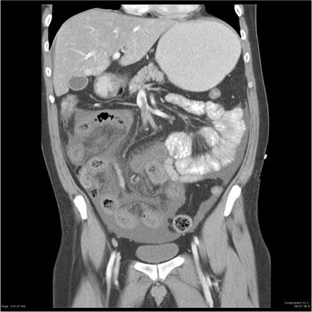
The splenic vein is dilated 103mm. Mesenteric venous thrombosis MVT describes acute subacute or chronic thrombosis of the superior or inferior mesenteric vein or branches. 12 Clinically there are two subtypes of mesenteric ischemia. Acute portal vein thrombosis PVT is characterized by the recent development of a thrombus in the portal vein or its left or right branches. Mesenteric vein thrombosis almost always involves the distal small intestine superior mesenteric venous drainage and rarely involves the colon inferior mesenteric venous drainage.
 Source: radiopaedia.org
Chronic mesenteric venous thrombosis is differentiated from acute mesenteric venous thrombosis by the existence of collateral venous circulation and cavernoma around the. Acute thrombosis commonly presents with abdominal pain and chronic type with features of portal hypertension. Often the superior mesenteric vein is considered the common trunk after all the chief tributaries have joined. CT chest abdomen and pelvis revealed an extensive thrombus extending from the portal vein to the superior mesenteric vein. The symptoms are usually not typical enough to offer a clinical diagnosis.
 Source: radiopaedia.org
The splenic vein is dilated 103mm. The splenic vein is dilated 103mm. A Contrast-enhanced maximum intensity projection magnetic resonance MR angiogram reveals collateral vessels from a superior mesenteric vein branch via submucosal varices into liver. In one retrospective study on 60 patients with chronic thrombosis of PVs or superior mesenteric veins 39 with variceal bleeding 18 with thrombophilia 9 with variceal bleeding received anticoagulation with recanalization of veins in 3 patients whereas none of the patients who were not anticoagulated recanalized the veins. Mesenteric vein thrombosis is increasingly recognized as a cause of mesenteric ischemia.
 Source: nejm.org
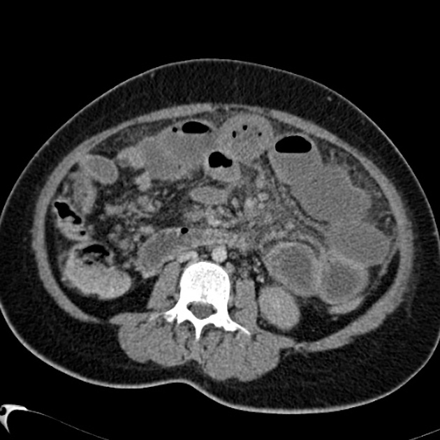
Chronic mesenteric venous thrombosis is differentiated from acute mesenteric venous thrombosis by the existence of collateral venous circulation and cavernoma around the. Non opacification of main trunk of superior mesenteric vein up to the portosplenic confluence with perivascular fat stranding. MVT may present with acute abdominal pain or may be an asymptomatic incidental finding on abdominal imaging. Spleen is enlarged spanning 20cm. In one retrospective study on 60 patients with chronic thrombosis of PVs or superior mesenteric veins 39 with variceal bleeding 18 with thrombophilia 9 with variceal bleeding received anticoagulation with recanalization of veins in 3 patients whereas none of the patients who were not anticoagulated recanalized the veins.
 Source: radiopaedia.org
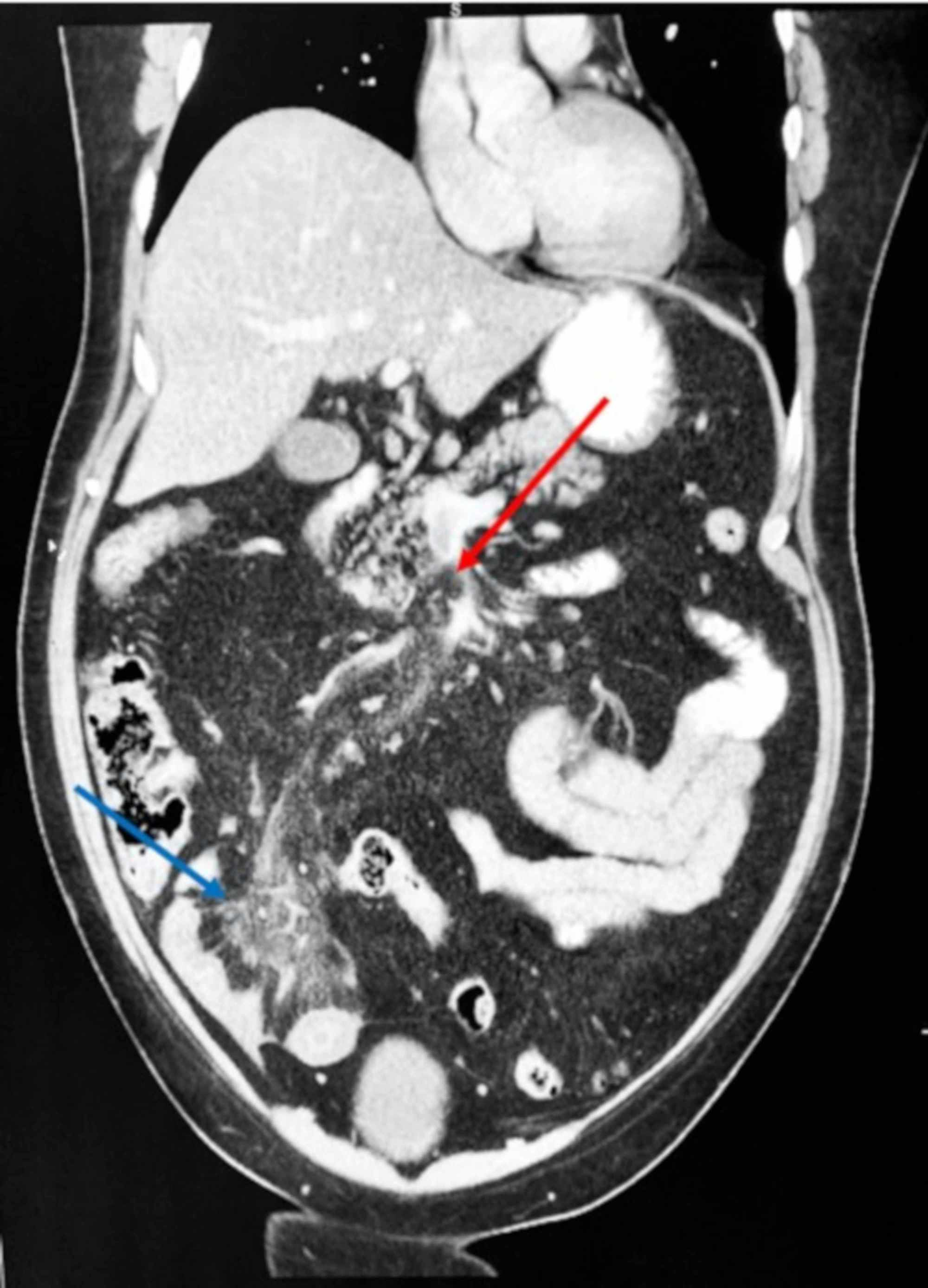
The diagnosis of mesenteric vein thrombosis relies heavily on imaging. MVT accounts for 1 in 5000 to 15000 inpa - tient admissions and 1 in 1000 emer-. We describe the computed tomography CT appearances of four patients with acute or acute on chronic case 3 pancreatitis which demonstrated isolated superior mesenteric vein SMV thrombosis. Contrast enhanced CT scan of abdomen is quite accurate for diagnosing and differentiating two types of mesenteric venous thrombosis. Mesenteric vein thrombosis is increasingly recognized as a cause of mesenteric ischemia.
 Source: radiopaedia.org
10 Further recurrent bleeding was. Mesenteric venous thrombosis MVT describes acute subacute or chronic thrombosis of the superior or inferior mesenteric vein or branches. Chronic mesenteric venous thrombosis accounts for approximately 20 to 40 of total mesenteric venous thrombosis cases and rarely causes intestinal infarction. 10 Further recurrent bleeding was. 12 Clinically there are two subtypes of mesenteric ischemia.
 Source: westjem.com
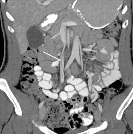
MVT may present with acute abdominal pain or may be an asymptomatic incidental finding on abdominal imaging. Arrow submucosal varices in biliary enteric anastomosis. Gross anatomy Origin and course. Main portal vein is dilated 145mm. In three of the four cases follow-up CT scans showed the SMV thrombosis to have.
This site is an open community for users to submit their favorite wallpapers on the internet, all images or pictures in this website are for personal wallpaper use only, it is stricly prohibited to use this wallpaper for commercial purposes, if you are the author and find this image is shared without your permission, please kindly raise a DMCA report to Us.
If you find this site helpful, please support us by sharing this posts to your favorite social media accounts like Facebook, Instagram and so on or you can also bookmark this blog page with the title chronic superior mesenteric vein thrombosis radiology by using Ctrl + D for devices a laptop with a Windows operating system or Command + D for laptops with an Apple operating system. If you use a smartphone, you can also use the drawer menu of the browser you are using. Whether it’s a Windows, Mac, iOS or Android operating system, you will still be able to bookmark this website.


