Your Inferior mesenteric artery occlusion ct images are available. Inferior mesenteric artery occlusion ct are a topic that is being searched for and liked by netizens now. You can Download the Inferior mesenteric artery occlusion ct files here. Get all free photos.
If you’re looking for inferior mesenteric artery occlusion ct pictures information related to the inferior mesenteric artery occlusion ct interest, you have visit the ideal site. Our site frequently gives you hints for refferencing the highest quality video and picture content, please kindly search and find more enlightening video content and graphics that fit your interests.
Inferior Mesenteric Artery Occlusion Ct. 28 Superior mesenteric vein thrombosis has historically been difficult to diagnose. Celiac axis superior mesenteric artery and inferior mesenteric artery occlusion. On the basis of our personal experience with thousands of abdomi-nal CTA and MRA examinations the inferior mesenteric artery and th e periphery of the other splanchnic vessels are currently better assessed. 2 The use of water as an oral contrast agent allows for improved visualization of the bowel wall and mucosa.
 Inferior Mesenteric Artery Aneurysm Radiology Case Radiopaedia Org From radiopaedia.org
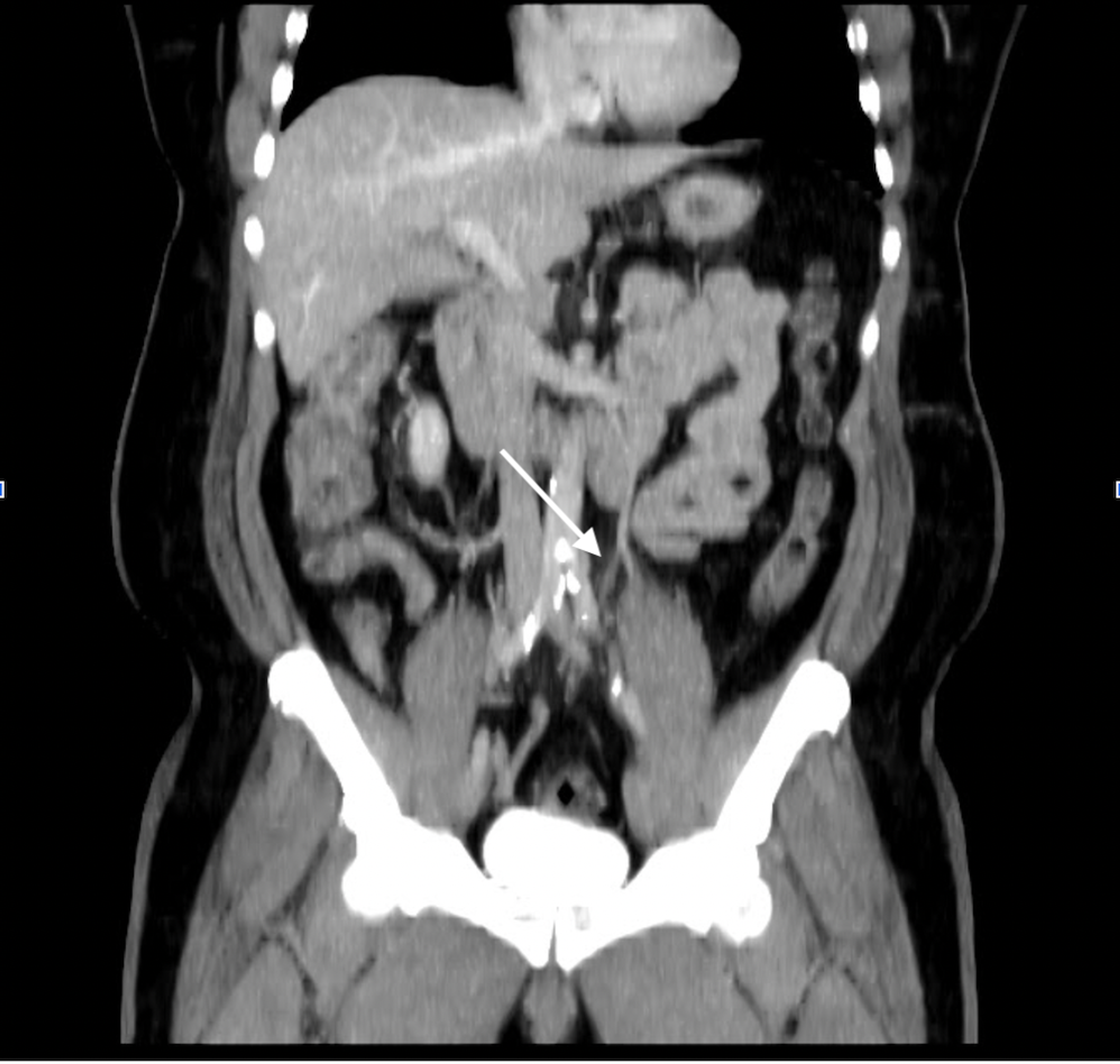
Features are consistent with inferior mesenteric artery IMA ischemic colitis. A detailed description of mesenteric artery thrombosis is outside the scope of this chapter but is provided in other sources. K551 is a billablespecific ICD-10-CM code that can be used to indicate a diagnosis for reimbursement purposes. It is a severe and potentially fatal illness typically of the superior mesenteric artery SMA which provides the primary arterial supply to the small intestine and ascending colon. The occlusion may occur due to in-situ thrombosis of the. To demonstrate the clinical applicability of 3-dimensional CT angiography 3D-CTA in evaluating the anatomic variations of inferior mesenteric artery IMA and left colic artery LCA to help make pre-operative strategies of rectal cancer surgery.
On contrast-enhanced CT thrombus in the mesenteric and portal veins is usually visible and mesenteric venous obstruction can be confirmed by CT in more than 90 of cases 28 30 Fig.
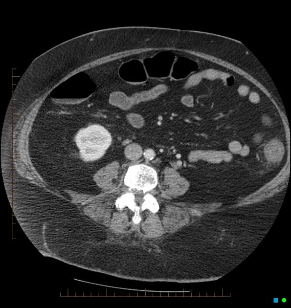
Factors that predispose patients to atherosclerosis. Venous causes of acute mesenteric ischemia are less common 515 of cases 4 9 and are most often the result of a thrombosis of the. Celiac axis superior mesenteric artery and inferior mesenteric artery occlusion. Mesenteric artery thrombosis MAT is a condition involving occlusion of the arterial vascular supply of the intestinal system. On the basis of our personal experience with thousands of abdomi-nal CTA and MRA examinations the inferior mesenteric artery and th e periphery of the other splanchnic vessels are currently better assessed. CT image demonstrating the inferior mesenteric artery aneurysm.
 Source: cureus.com
2 The use of water as an oral contrast agent allows for improved visualization of the bowel wall and mucosa. Return from the CT examination the patient experienced intense abdominal pain associated with nausea. It also contributes to the formation of the marginal artery of Drummond. Factors that predispose patients to atherosclerosis. The 2022 edition of ICD-10-CM K551 became effective on October 1 2021.
 Source: researchgate.net
Mesenteric artery thrombosis MAT is a condition involving occlusion of the arterial vascular supply of the intestinal system. K551 is a billablespecific ICD-10-CM code that can be used to indicate a diagnosis for reimbursement purposes. Venous causes of acute mesenteric ischemia are less common 515 of cases 4 9 and are most often the result of a thrombosis of the. Contrast-enhanced computed tomography CT and aortography revealed thoracoabdominal aortic aneurysm of 6 cm in diameter accompanied by inferior mesenteric aneurysm of 3 cm in diameter. The aneurysm was further assessed by angiography which demonstrated complete occlusion of the superior mesenteric artery SMA coeliac trunk and right renal artery.
 Source: semanticscholar.org
3 Acquiring images in the arterial and venous phases biphasic allows for evaluating the arterial and venous mesenteric vessels as. It supplies the hindgut and has four major branches called left colic sigmoid and superior rectal arteries. However it still supplied perfusion to most of the collaterals in lower extremities Figure 1. At these sites stenosis or occlusion of the lumen is observed at contrast-enhanced CT and is well appreciated on sagittal images and reformatted angiograms Fig 11. In the acute phase the presence of pericolic fluid was found in 100 of patients undergoing progressive resorption.
 Source: researchgate.net
In the acute phase the presence of pericolic fluid was found in 100 of patients undergoing progressive resorption. However it still supplied perfusion to most of the collaterals in lower extremities Figure 1. A CT angiography of the aorta and lower extremities was ordered to better evaluate the distal circulation. A detailed description of mesenteric artery thrombosis is outside the scope of this chapter but is provided in other sources. Computed tomography has been heralded as the primary imaging for the evaluation of mesenteric ischemia.
 Source: researchgate.net
In the acute phase the presence of pericolic fluid was found in 100 of patients undergoing progressive resorption. The inferior mesenteric artery is opacified by contrast only for the first centimeter or so whereupon it reduces in caliber. Features are consistent with inferior mesenteric artery IMA ischemic colitis. Computed tomography has been heralded as the primary imaging for the evaluation of mesenteric ischemia. Overall SMA occlusion due to emboli or thrombi is responsible for 6070 of cases of acute mesenteric ischemia whereas nonocclusive mesenteric ischemia accounts for 2030 of cases 910.
 Source: researchgate.net
To demonstrate the clinical applicability of 3-dimensional CT angiography 3D-CTA in evaluating the anatomic variations of inferior mesenteric artery IMA and left colic artery LCA to help make pre-operative strategies of rectal cancer surgery. On contrast-enhanced CT thrombus in the mesenteric and portal veins is usually visible and mesenteric venous obstruction can be confirmed by CT in more than 90 of cases 28 30 Fig. The inferior mesenteric artery is the last of the three major anterior branches of the abdominal aorta the other two are the coeliac trunk and superior mesenteric artery. The occlusion may occur due to in-situ thrombosis of the. The inferior mesenteric artery is opacified by contrast only for the first centimeter or so whereupon it reduces in caliber.
 Source: jem-journal.com
2 The use of water as an oral contrast agent allows for improved visualization of the bowel wall and mucosa. A detailed description of mesenteric artery thrombosis is outside the scope of this chapter but is provided in other sources. A CT angiography of the aorta and lower extremities was ordered to better evaluate the distal circulation. The etiology of chronic mesenteric ischemia is often multifactorial. On the basis of our personal experience with thousands of abdomi-nal CTA and MRA examinations the inferior mesenteric artery and th e periphery of the other splanchnic vessels are currently better assessed.
 Source: clinicalradiologyonline.net
Computed tomography has been heralded as the primary imaging for the evaluation of mesenteric ischemia. Among the 32 CT examinations performed in the acute phase 625 did not present signs of occlusion of the superior mesenteric artery SMA or inferior mesenteric artery IMA whereas IMA occlusion was detected in 375 of CT examinations. Venous causes of acute mesenteric ischemia are less common 515 of cases 4 9 and are most often the result of a thrombosis of the. 3 Acquiring images in the arterial and venous phases biphasic allows for evaluating the arterial and venous mesenteric vessels as. CT angiography showed an occlusion at the origin of the coeliac trunk CTr and superior mesenteric artery SMA a focal stenosis 50 at the origin of the inferior mesenteric artery IMA and a hypertrophy of Riolanos arch with rehabitation of SMA and CTr confirming the diagnosis of abdominal claudication Fig.
 Source: radiopaedia.org
A detailed description of mesenteric artery thrombosis is outside the scope of this chapter but is provided in other sources. Factors that predispose patients to atherosclerosis. 1 and also an occlusion of the right popliteal artery were evident on CT. 283132 Compared with mesenteric artery occlusion thrombosis of the superior and inferior mesenteric veins is less common and less precipitous. The 2022 edition of ICD-10-CM K551 became effective on October 1 2021.
 Source: researchgate.net
Proximal or segmental mesenteric artery steno-sis or occlusion in only one affected vessel is rare but can occur Fig. Distal branches of vein are engorged. 283132 Compared with mesenteric artery occlusion thrombosis of the superior and inferior mesenteric veins is less common and less precipitous. This is the American ICD-10-CM version of K551 - other international versions of ICD-10 K551 may differ. 1 and also an occlusion of the right popliteal artery were evident on CT.
 Source: radiopaedia.org
However it still supplied perfusion to most of the collaterals in lower extremities Figure 1. On the basis of our personal experience with thousands of abdomi-nal CTA and MRA examinations the inferior mesenteric artery and th e periphery of the other splanchnic vessels are currently better assessed. CT angiography showed an occlusion at the origin of the coeliac trunk CTr and superior mesenteric artery SMA a focal stenosis 50 at the origin of the inferior mesenteric artery IMA and a hypertrophy of Riolanos arch with rehabitation of SMA and CTr confirming the diagnosis of abdominal claudication Fig. Crawford ES Morris GC Jr Myhre HO Roehm JO Jr Surgery 1977 Dec826856-66. At these sites stenosis or occlusion of the lumen is observed at contrast-enhanced CT and is well appreciated on sagittal images and reformatted angiograms Fig 11.
 Source: researchgate.net
This is the American ICD-10-CM version of K551 - other international versions of ICD-10 K551 may differ. In the acute phase the presence of pericolic fluid was found in 100 of patients undergoing progressive resorption. 3 Less common etiologies include dissection vasculitis fibromuscular dysplasia radiation and cocaine abuse. This is the American ICD-10-CM version of K551 - other international versions of ICD-10 K551 may differ. A CT angiography of the aorta and lower extremities was ordered to better evaluate the distal circulation.
 Source: ejves.com
A CT angiography of the aorta and lower extremities was ordered to better evaluate the distal circulation. The 2022 edition of ICD-10-CM K551 became effective on October 1 2021. The IMA aneurysm was delineated and also ostial stenosis of the same vessel. The most common cause is atherosclerosis involving the proximal portions of the celiac superior mesenteric or inferior mesenteric artery. 3 Less common etiologies include dissection vasculitis fibromuscular dysplasia radiation and cocaine abuse.
 Source: researchgate.net
The occlusion may occur due to in-situ thrombosis of the. CT image demonstrating the inferior mesenteric artery aneurysm. The most common cause is atherosclerosis involving the proximal portions of the celiac superior mesenteric or inferior mesenteric artery. Crawford ES Morris GC Jr Myhre HO Roehm JO Jr Surgery 1977 Dec826856-66. Worldwide there have been only eleven reports of IMA aneurysms associated with tight stenosis of the SMA and celiac trunk 5 6.
 Source: aneskey.com
To demonstrate the clinical applicability of 3-dimensional CT angiography 3D-CTA in evaluating the anatomic variations of inferior mesenteric artery IMA and left colic artery LCA to help make pre-operative strategies of rectal cancer surgery. The aneurysm was further assessed by angiography which demonstrated complete occlusion of the superior mesenteric artery SMA coeliac trunk and right renal artery. Severe calcification of the abdominal aorta and occlusion of the celiac and the superior mesenteric arteries were also noted whose territories were perfused by collateral circulation. Worldwide there have been only eleven reports of IMA aneurysms associated with tight stenosis of the SMA and celiac trunk 5 6. The etiology of chronic mesenteric ischemia is often multifactorial.

The most common cause is atherosclerosis involving the proximal portions of the celiac superior mesenteric or inferior mesenteric artery. On contrast-enhanced CT thrombus in the mesenteric and portal veins is usually visible and mesenteric venous obstruction can be confirmed by CT in more than 90 of cases 28 30 Fig. Among the 32 CT examinations performed in the acute phase 625 did not present signs of occlusion of the superior mesenteric artery SMA or inferior mesenteric artery IMA whereas IMA occlusion was detected in 375 of CT examinations. Venous causes of acute mesenteric ischemia are less common 515 of cases 4 9 and are most often the result of a thrombosis of the. At these sites stenosis or occlusion of the lumen is observed at contrast-enhanced CT and is well appreciated on sagittal images and reformatted angiograms Fig 11.
 Source: researchgate.net
Worldwide there have been only eleven reports of IMA aneurysms associated with tight stenosis of the SMA and celiac trunk 5 6. At these sites stenosis or occlusion of the lumen is observed at contrast-enhanced CT and is well appreciated on sagittal images and reformatted angiograms Fig 11. In the acute phase the presence of pericolic fluid was found in 100 of patients undergoing progressive resorption. Contrast-enhanced computed tomography CT and aortography revealed thoracoabdominal aortic aneurysm of 6 cm in diameter accompanied by inferior mesenteric aneurysm of 3 cm in diameter. Celiac axis superior mesenteric artery and inferior mesenteric artery occlusion.
 Source: radiopaedia.org
In the acute phase the presence of pericolic fluid was found in 100 of patients undergoing progressive resorption. At these sites stenosis or occlusion of the lumen is observed at contrast-enhanced CT and is well appreciated on sagittal images and reformatted angiograms Fig 11. Return from the CT examination the patient experienced intense abdominal pain associated with nausea. 188 patients with abdominal and pelvic contrast-enhanced CT scan were retrospectively enrolled and 3D-CTA. Mesenteric artery thrombosis MAT is a condition involving occlusion of the arterial vascular supply of the intestinal system.
This site is an open community for users to do sharing their favorite wallpapers on the internet, all images or pictures in this website are for personal wallpaper use only, it is stricly prohibited to use this wallpaper for commercial purposes, if you are the author and find this image is shared without your permission, please kindly raise a DMCA report to Us.
If you find this site beneficial, please support us by sharing this posts to your own social media accounts like Facebook, Instagram and so on or you can also save this blog page with the title inferior mesenteric artery occlusion ct by using Ctrl + D for devices a laptop with a Windows operating system or Command + D for laptops with an Apple operating system. If you use a smartphone, you can also use the drawer menu of the browser you are using. Whether it’s a Windows, Mac, iOS or Android operating system, you will still be able to bookmark this website.


