Your Inferior mesenteric vein anatomy images are available. Inferior mesenteric vein anatomy are a topic that is being searched for and liked by netizens today. You can Get the Inferior mesenteric vein anatomy files here. Get all royalty-free photos.
If you’re looking for inferior mesenteric vein anatomy pictures information connected with to the inferior mesenteric vein anatomy keyword, you have visit the right site. Our website frequently provides you with suggestions for seeing the highest quality video and picture content, please kindly hunt and find more informative video content and images that fit your interests.
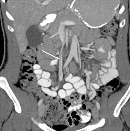
Inferior Mesenteric Vein Anatomy. In most individuals the portal vein is formed by the union of the superior mesenteric vein and the splenic vein. For this reason the portal vein is occasionally called the splenic-mesenteric confluence. The inferior vena cava is a large vein that carries the deoxygenated blood from the lower and middle body into the right atrium of the heartIt is formed by the joining of the right and the left common iliac veins usually at the level of the fifth lumbar vertebra. Measuring approximately 8 cm 3 inches long in adults the portal vein is located in the right upper quadrant of the abdomen originating behind the neck of the pancreas.
 Variations And Anomalies Of Hepatic Portal Vein Anatomy Cystic Vein Portal Vein 1 09 Cm Superior Pancreaticoduoden Anatomy Veins Anatomy And Physiology From pinterest.com
In most individuals the portal vein is formed by the union of the superior mesenteric vein and the splenic vein. The inferior vena cava is the lower inferior of the two venae cavae the two large veins that carry deoxygenated blood. Measuring approximately 8 cm 3 inches long in adults the portal vein is located in the right upper quadrant of the abdomen originating behind the neck of the pancreas. For this reason the portal vein is occasionally called the splenic-mesenteric confluence. The inferior vena cava is a large vein that carries the deoxygenated blood from the lower and middle body into the right atrium of the heartIt is formed by the joining of the right and the left common iliac veins usually at the level of the fifth lumbar vertebra. On normal anatomy typically the splenic vein SV joins the superior mesenteric vein SMV anteriorly to the IVC and posteriorly to the pancreatic neck to form the PV which ascends within the hepatoduodenal ligament posteriorly to the hepatic artery and common bile duct toward the hepatic hilum where it divides into right and left Fig.
The inferior vena cava is a large vein that carries the deoxygenated blood from the lower and middle body into the right atrium of the heartIt is formed by the joining of the right and the left common iliac veins usually at the level of the fifth lumbar vertebra.
For this reason the portal vein is occasionally called the splenic-mesenteric confluence. Measuring approximately 8 cm 3 inches long in adults the portal vein is located in the right upper quadrant of the abdomen originating behind the neck of the pancreas. The inferior vena cava is a large vein that carries the deoxygenated blood from the lower and middle body into the right atrium of the heartIt is formed by the joining of the right and the left common iliac veins usually at the level of the fifth lumbar vertebra. The inferior vena cava is the lower inferior of the two venae cavae the two large veins that carry deoxygenated blood. In most individuals the portal vein is formed by the union of the superior mesenteric vein and the splenic vein. On normal anatomy typically the splenic vein SV joins the superior mesenteric vein SMV anteriorly to the IVC and posteriorly to the pancreatic neck to form the PV which ascends within the hepatoduodenal ligament posteriorly to the hepatic artery and common bile duct toward the hepatic hilum where it divides into right and left Fig.
Source: pinterest.com
Measuring approximately 8 cm 3 inches long in adults the portal vein is located in the right upper quadrant of the abdomen originating behind the neck of the pancreas. The inferior vena cava is a large vein that carries the deoxygenated blood from the lower and middle body into the right atrium of the heartIt is formed by the joining of the right and the left common iliac veins usually at the level of the fifth lumbar vertebra. In most individuals the portal vein is formed by the union of the superior mesenteric vein and the splenic vein. For this reason the portal vein is occasionally called the splenic-mesenteric confluence. The inferior vena cava is the lower inferior of the two venae cavae the two large veins that carry deoxygenated blood.
 Source: pinterest.com
The inferior vena cava is the lower inferior of the two venae cavae the two large veins that carry deoxygenated blood. The inferior vena cava is the lower inferior of the two venae cavae the two large veins that carry deoxygenated blood. For this reason the portal vein is occasionally called the splenic-mesenteric confluence. The inferior vena cava is a large vein that carries the deoxygenated blood from the lower and middle body into the right atrium of the heartIt is formed by the joining of the right and the left common iliac veins usually at the level of the fifth lumbar vertebra. Measuring approximately 8 cm 3 inches long in adults the portal vein is located in the right upper quadrant of the abdomen originating behind the neck of the pancreas.
 Source: pinterest.com
Measuring approximately 8 cm 3 inches long in adults the portal vein is located in the right upper quadrant of the abdomen originating behind the neck of the pancreas. The inferior vena cava is the lower inferior of the two venae cavae the two large veins that carry deoxygenated blood. In most individuals the portal vein is formed by the union of the superior mesenteric vein and the splenic vein. For this reason the portal vein is occasionally called the splenic-mesenteric confluence. The inferior vena cava is a large vein that carries the deoxygenated blood from the lower and middle body into the right atrium of the heartIt is formed by the joining of the right and the left common iliac veins usually at the level of the fifth lumbar vertebra.
 Source: pinterest.com
The inferior vena cava is a large vein that carries the deoxygenated blood from the lower and middle body into the right atrium of the heartIt is formed by the joining of the right and the left common iliac veins usually at the level of the fifth lumbar vertebra. Measuring approximately 8 cm 3 inches long in adults the portal vein is located in the right upper quadrant of the abdomen originating behind the neck of the pancreas. The inferior vena cava is the lower inferior of the two venae cavae the two large veins that carry deoxygenated blood. In most individuals the portal vein is formed by the union of the superior mesenteric vein and the splenic vein. On normal anatomy typically the splenic vein SV joins the superior mesenteric vein SMV anteriorly to the IVC and posteriorly to the pancreatic neck to form the PV which ascends within the hepatoduodenal ligament posteriorly to the hepatic artery and common bile duct toward the hepatic hilum where it divides into right and left Fig.
 Source: pinterest.com
The inferior vena cava is the lower inferior of the two venae cavae the two large veins that carry deoxygenated blood. In most individuals the portal vein is formed by the union of the superior mesenteric vein and the splenic vein. The inferior vena cava is a large vein that carries the deoxygenated blood from the lower and middle body into the right atrium of the heartIt is formed by the joining of the right and the left common iliac veins usually at the level of the fifth lumbar vertebra. Measuring approximately 8 cm 3 inches long in adults the portal vein is located in the right upper quadrant of the abdomen originating behind the neck of the pancreas. The inferior vena cava is the lower inferior of the two venae cavae the two large veins that carry deoxygenated blood.
 Source: pinterest.com
For this reason the portal vein is occasionally called the splenic-mesenteric confluence. Measuring approximately 8 cm 3 inches long in adults the portal vein is located in the right upper quadrant of the abdomen originating behind the neck of the pancreas. The inferior vena cava is the lower inferior of the two venae cavae the two large veins that carry deoxygenated blood. For this reason the portal vein is occasionally called the splenic-mesenteric confluence. In most individuals the portal vein is formed by the union of the superior mesenteric vein and the splenic vein.
 Source: pinterest.com
Measuring approximately 8 cm 3 inches long in adults the portal vein is located in the right upper quadrant of the abdomen originating behind the neck of the pancreas. For this reason the portal vein is occasionally called the splenic-mesenteric confluence. On normal anatomy typically the splenic vein SV joins the superior mesenteric vein SMV anteriorly to the IVC and posteriorly to the pancreatic neck to form the PV which ascends within the hepatoduodenal ligament posteriorly to the hepatic artery and common bile duct toward the hepatic hilum where it divides into right and left Fig. The inferior vena cava is the lower inferior of the two venae cavae the two large veins that carry deoxygenated blood. Measuring approximately 8 cm 3 inches long in adults the portal vein is located in the right upper quadrant of the abdomen originating behind the neck of the pancreas.
 Source: pinterest.com
On normal anatomy typically the splenic vein SV joins the superior mesenteric vein SMV anteriorly to the IVC and posteriorly to the pancreatic neck to form the PV which ascends within the hepatoduodenal ligament posteriorly to the hepatic artery and common bile duct toward the hepatic hilum where it divides into right and left Fig. On normal anatomy typically the splenic vein SV joins the superior mesenteric vein SMV anteriorly to the IVC and posteriorly to the pancreatic neck to form the PV which ascends within the hepatoduodenal ligament posteriorly to the hepatic artery and common bile duct toward the hepatic hilum where it divides into right and left Fig. In most individuals the portal vein is formed by the union of the superior mesenteric vein and the splenic vein. Measuring approximately 8 cm 3 inches long in adults the portal vein is located in the right upper quadrant of the abdomen originating behind the neck of the pancreas. For this reason the portal vein is occasionally called the splenic-mesenteric confluence.
 Source: pinterest.com
The inferior vena cava is a large vein that carries the deoxygenated blood from the lower and middle body into the right atrium of the heartIt is formed by the joining of the right and the left common iliac veins usually at the level of the fifth lumbar vertebra. The inferior vena cava is a large vein that carries the deoxygenated blood from the lower and middle body into the right atrium of the heartIt is formed by the joining of the right and the left common iliac veins usually at the level of the fifth lumbar vertebra. The inferior vena cava is the lower inferior of the two venae cavae the two large veins that carry deoxygenated blood. For this reason the portal vein is occasionally called the splenic-mesenteric confluence. On normal anatomy typically the splenic vein SV joins the superior mesenteric vein SMV anteriorly to the IVC and posteriorly to the pancreatic neck to form the PV which ascends within the hepatoduodenal ligament posteriorly to the hepatic artery and common bile duct toward the hepatic hilum where it divides into right and left Fig.
 Source: cz.pinterest.com
On normal anatomy typically the splenic vein SV joins the superior mesenteric vein SMV anteriorly to the IVC and posteriorly to the pancreatic neck to form the PV which ascends within the hepatoduodenal ligament posteriorly to the hepatic artery and common bile duct toward the hepatic hilum where it divides into right and left Fig. Measuring approximately 8 cm 3 inches long in adults the portal vein is located in the right upper quadrant of the abdomen originating behind the neck of the pancreas. The inferior vena cava is a large vein that carries the deoxygenated blood from the lower and middle body into the right atrium of the heartIt is formed by the joining of the right and the left common iliac veins usually at the level of the fifth lumbar vertebra. The inferior vena cava is the lower inferior of the two venae cavae the two large veins that carry deoxygenated blood. In most individuals the portal vein is formed by the union of the superior mesenteric vein and the splenic vein.
 Source: pinterest.com
The inferior vena cava is a large vein that carries the deoxygenated blood from the lower and middle body into the right atrium of the heartIt is formed by the joining of the right and the left common iliac veins usually at the level of the fifth lumbar vertebra. In most individuals the portal vein is formed by the union of the superior mesenteric vein and the splenic vein. On normal anatomy typically the splenic vein SV joins the superior mesenteric vein SMV anteriorly to the IVC and posteriorly to the pancreatic neck to form the PV which ascends within the hepatoduodenal ligament posteriorly to the hepatic artery and common bile duct toward the hepatic hilum where it divides into right and left Fig. The inferior vena cava is the lower inferior of the two venae cavae the two large veins that carry deoxygenated blood. Measuring approximately 8 cm 3 inches long in adults the portal vein is located in the right upper quadrant of the abdomen originating behind the neck of the pancreas.
 Source: pinterest.com
Measuring approximately 8 cm 3 inches long in adults the portal vein is located in the right upper quadrant of the abdomen originating behind the neck of the pancreas. For this reason the portal vein is occasionally called the splenic-mesenteric confluence. The inferior vena cava is a large vein that carries the deoxygenated blood from the lower and middle body into the right atrium of the heartIt is formed by the joining of the right and the left common iliac veins usually at the level of the fifth lumbar vertebra. Measuring approximately 8 cm 3 inches long in adults the portal vein is located in the right upper quadrant of the abdomen originating behind the neck of the pancreas. On normal anatomy typically the splenic vein SV joins the superior mesenteric vein SMV anteriorly to the IVC and posteriorly to the pancreatic neck to form the PV which ascends within the hepatoduodenal ligament posteriorly to the hepatic artery and common bile duct toward the hepatic hilum where it divides into right and left Fig.
 Source: pinterest.com
The inferior vena cava is a large vein that carries the deoxygenated blood from the lower and middle body into the right atrium of the heartIt is formed by the joining of the right and the left common iliac veins usually at the level of the fifth lumbar vertebra. For this reason the portal vein is occasionally called the splenic-mesenteric confluence. In most individuals the portal vein is formed by the union of the superior mesenteric vein and the splenic vein. The inferior vena cava is the lower inferior of the two venae cavae the two large veins that carry deoxygenated blood. On normal anatomy typically the splenic vein SV joins the superior mesenteric vein SMV anteriorly to the IVC and posteriorly to the pancreatic neck to form the PV which ascends within the hepatoduodenal ligament posteriorly to the hepatic artery and common bile duct toward the hepatic hilum where it divides into right and left Fig.
 Source: pinterest.com
The inferior vena cava is the lower inferior of the two venae cavae the two large veins that carry deoxygenated blood. In most individuals the portal vein is formed by the union of the superior mesenteric vein and the splenic vein. For this reason the portal vein is occasionally called the splenic-mesenteric confluence. The inferior vena cava is the lower inferior of the two venae cavae the two large veins that carry deoxygenated blood. Measuring approximately 8 cm 3 inches long in adults the portal vein is located in the right upper quadrant of the abdomen originating behind the neck of the pancreas.
 Source: pinterest.com
In most individuals the portal vein is formed by the union of the superior mesenteric vein and the splenic vein. For this reason the portal vein is occasionally called the splenic-mesenteric confluence. Measuring approximately 8 cm 3 inches long in adults the portal vein is located in the right upper quadrant of the abdomen originating behind the neck of the pancreas. In most individuals the portal vein is formed by the union of the superior mesenteric vein and the splenic vein. On normal anatomy typically the splenic vein SV joins the superior mesenteric vein SMV anteriorly to the IVC and posteriorly to the pancreatic neck to form the PV which ascends within the hepatoduodenal ligament posteriorly to the hepatic artery and common bile duct toward the hepatic hilum where it divides into right and left Fig.
 Source: pinterest.com
Measuring approximately 8 cm 3 inches long in adults the portal vein is located in the right upper quadrant of the abdomen originating behind the neck of the pancreas. The inferior vena cava is a large vein that carries the deoxygenated blood from the lower and middle body into the right atrium of the heartIt is formed by the joining of the right and the left common iliac veins usually at the level of the fifth lumbar vertebra. On normal anatomy typically the splenic vein SV joins the superior mesenteric vein SMV anteriorly to the IVC and posteriorly to the pancreatic neck to form the PV which ascends within the hepatoduodenal ligament posteriorly to the hepatic artery and common bile duct toward the hepatic hilum where it divides into right and left Fig. The inferior vena cava is the lower inferior of the two venae cavae the two large veins that carry deoxygenated blood. In most individuals the portal vein is formed by the union of the superior mesenteric vein and the splenic vein.
 Source: pinterest.com
The inferior vena cava is a large vein that carries the deoxygenated blood from the lower and middle body into the right atrium of the heartIt is formed by the joining of the right and the left common iliac veins usually at the level of the fifth lumbar vertebra. In most individuals the portal vein is formed by the union of the superior mesenteric vein and the splenic vein. Measuring approximately 8 cm 3 inches long in adults the portal vein is located in the right upper quadrant of the abdomen originating behind the neck of the pancreas. On normal anatomy typically the splenic vein SV joins the superior mesenteric vein SMV anteriorly to the IVC and posteriorly to the pancreatic neck to form the PV which ascends within the hepatoduodenal ligament posteriorly to the hepatic artery and common bile duct toward the hepatic hilum where it divides into right and left Fig. The inferior vena cava is the lower inferior of the two venae cavae the two large veins that carry deoxygenated blood.
 Source: pinterest.com
The inferior vena cava is a large vein that carries the deoxygenated blood from the lower and middle body into the right atrium of the heartIt is formed by the joining of the right and the left common iliac veins usually at the level of the fifth lumbar vertebra. In most individuals the portal vein is formed by the union of the superior mesenteric vein and the splenic vein. For this reason the portal vein is occasionally called the splenic-mesenteric confluence. The inferior vena cava is a large vein that carries the deoxygenated blood from the lower and middle body into the right atrium of the heartIt is formed by the joining of the right and the left common iliac veins usually at the level of the fifth lumbar vertebra. Measuring approximately 8 cm 3 inches long in adults the portal vein is located in the right upper quadrant of the abdomen originating behind the neck of the pancreas.
This site is an open community for users to do submittion their favorite wallpapers on the internet, all images or pictures in this website are for personal wallpaper use only, it is stricly prohibited to use this wallpaper for commercial purposes, if you are the author and find this image is shared without your permission, please kindly raise a DMCA report to Us.
If you find this site good, please support us by sharing this posts to your own social media accounts like Facebook, Instagram and so on or you can also save this blog page with the title inferior mesenteric vein anatomy by using Ctrl + D for devices a laptop with a Windows operating system or Command + D for laptops with an Apple operating system. If you use a smartphone, you can also use the drawer menu of the browser you are using. Whether it’s a Windows, Mac, iOS or Android operating system, you will still be able to bookmark this website.