Your Inferior mesenteric vein thrombosis ct images are available in this site. Inferior mesenteric vein thrombosis ct are a topic that is being searched for and liked by netizens today. You can Download the Inferior mesenteric vein thrombosis ct files here. Get all free photos.
If you’re searching for inferior mesenteric vein thrombosis ct pictures information linked to the inferior mesenteric vein thrombosis ct interest, you have visit the ideal site. Our website always provides you with hints for downloading the maximum quality video and picture content, please kindly surf and locate more enlightening video articles and images that fit your interests.
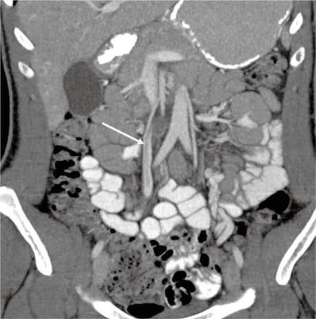
Inferior Mesenteric Vein Thrombosis Ct. Overview Mesenteric venous thrombosis MVT describes acute subacute or chronic thrombosis of the superior or inferior mesenteric vein or branches. Inflammatory changes around the sigmoid colon with a hyperdense diverticulum were also noted on CT scan Figure 4 suggestive of diverticulitis. Mild symptoms of covid-19 complicated by inferior mesenteric vein thrombosis. The superior mesenteric vein is most commonly involved.
 Abdominal Ct Findings Six Weeks Later Coronal Ct Image Showed Download Scientific Diagram From researchgate.net
Although mesenteric venous thrombosis is a relatively rare condition mortality remains high due to nonspecific symptoms. Mesenteric venous thrombosis is an uncommon but potentially lethal cause of bowel ischemia. MVT is a clot that blocks blood flow in a mesenteric vein. A computed tomography CT scan with intravenous contrast confirmed a thrombus in the inferior mesenteric vein IMV Figure 2 extending to the confluence of the IMV with the splenic vein Figure 3. It can be either acute presenting commonly with abdominal pain or chronic presenting with features of portal hypertension. There was also a report that 11.
There was also a report that 11.
The superior mesenteric vein is most commonly involved. CT of abdomen and pelvis was done with intravenous contrast and showed completely occluding continuous filling defect in the inferior mesenteric vein along its whole length up to portosplenic confluence extending slightly to. Primary MVT is idiopathic whereas secondary MVT can result. There was also a report that 11. It can be either acute presenting commonly with abdominal pain or chronic presenting with features of portal hypertension. Although mesenteric venous thrombosis is a relatively rare condition mortality remains high due to nonspecific symptoms.
 Source: westjem.com
MVT may present with acute abdominal pain. Mesenteric venous thrombosis MVT is a blood clot in one or more of the major veins that drain blood from the intestine. Mild symptoms of covid-19 complicated by inferior mesenteric vein thrombosis. Inflammatory changes around the sigmoid colon with a hyperdense diverticulum were also noted on CT scan Figure 4 suggestive of diverticulitis. Although mesenteric venous thrombosis is a relatively rare condition mortality remains high due to nonspecific symptoms.
 Source: radiopaedia.org
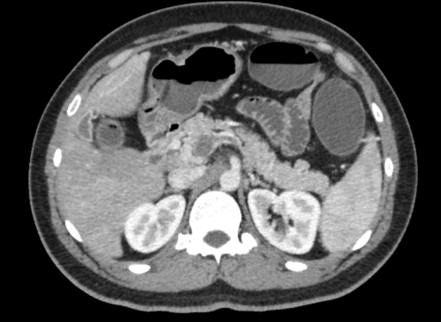
CT scan of the abdomen and pelvis which demonstrated thrombosis of the superior mesenteric vein Figure 1. CT diagnosis Abstract Mesenteric vein thrombosis has been described in association with such risk factors as coagulation disorders postoperative dehydration sepsis and trauma. Clinical symptoms are non-specific and include fevers abdominal pain and nausea 6. CASE DESCRIPTION A 33-year-old obese patient BMI 327 without other comorbidities was admitted to our hospital with complaints of severe low back pain radiated to the hypogastric region which had started about 8 hours before admission. Delayed detection or treatment of mesenteric venous thrombosis allows intestinal infarction to develop which can be life-threatening.
 Source: radiopaedia.org
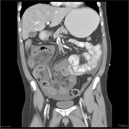
There was also a report that 11. CT Axial C portal venous phase There is evidence of superior mesenteric vein and portal vein thrombosis causing proximal small bowel dilatation and wall thickening with poor enhancement of dilated small bowel loops with suspicious pneumatosis intestinalis. There was also a report that 11. Doppler ultrasonography allows direct evaluation of the mesenteric and portal veins provides semiquantitative flow information and allows Doppler. CT of abdomen and pelvis was done with intravenous contrast and showed completely occluding continuous filling defect in the inferior mesenteric vein along its whole length up to portosplenic confluence extending slightly to.
 Source: radiopaedia.org
The management of mesenteric vein thrombosis. Open in a separate window. CASE DESCRIPTION A 33-year-old obese patient BMI 327 without other comorbidities was admitted to our hospital with complaints of severe low back pain radiated to the hypogastric region which had started about 8 hours before admission. The condition stops the blood circulation of the. The superior mesenteric vein is most commonly involved.
 Source: clinicalradiologyonline.net
Mesenteric vein thrombosis MVT accounts for 515 of all mes- enteric ischemic events and is classified as either primary or second- ary. In three of the four cases follow-up CT scans showed the SMV thrombosis to have resolved with resolution of the underlying pancreatitis without anticoagulation or surgical. Inflammatory changes around the sigmoid colon with a hyperdense diverticulum were also noted on CT scan Figure 4 suggestive of diverticulitis. Abdominal CT scan showed an enlarged inferior mesenteric vein not completely filled by contrast associated with infiltration of the adjacent adipose planes thus denoting mesenteric thrombosis Figure 2. Primary MVT is idiopathic whereas secondary MVT can result.
 Source: researchgate.net
There was also a report that 11. Abdominal CT scan showed an enlarged inferior mesenteric vein not completely filled by contrast associated with infiltration of the adjacent adipose planes thus denoting mesenteric thrombosis Figure 2. Mild symptoms of covid-19 complicated by inferior mesenteric vein thrombosis. Primary MVT is idiopathic whereas secondary MVT can result. Doppler ultrasonography allows direct evaluation of the mesenteric and portal veins provides semiquantitative flow information and allows Doppler.
 Source: researchgate.net
Pylephlebitis or suppurative thrombosis of the portal mesenteric venous system is a rare complication of intra-abdominal inflammatory processes such as diverticulitis appendicitis pancreatitis and inflammatory bowel disease 1 2 3 4 5. Mild symptoms of covid-19 complicated by inferior mesenteric vein thrombosis. We describe the computed tomography CT appearances of four patients with acute or acute on chronic case 3 pancreatitis which demonstrated isolated superior mesenteric vein SMV thrombosis. Chest computed tomography CT showed infiltration in a peripheral ground-glass pattern affecting both lower lobes suggestive of viral pneumonia Figure 1. There was also a report that 11.
 Source: thelancet.com
Inflammatory changes around the sigmoid colon with a hyperdense diverticulum were also noted on CT scan Figure 4 suggestive of diverticulitis. CT scan of the abdomen and pelvis which demonstrated thrombosis of the superior mesenteric vein Figure 1. Several imaging methods are available for diagnosis each of which has advantages and disadvantages. Open in a separate window. Primary MVT is idiopathic whereas secondary MVT can result.
 Source: researchgate.net
Mesenteric venous thrombosis is an uncommon but potentially lethal cause of bowel ischemia. The inter-reader agreement on CT for secondary intestinal abnormalities is slightly lower than for diagnosing MVT 8. Several imaging methods are available for diagnosis each of which has advantages and disadvantages. Delayed detection or treatment of mesenteric venous thrombosis allows intestinal infarction to develop which can be life-threatening. The superior mesenteric vein is most commonly involved.
 Source: ejves.com
We describe the computed tomography CT appearances of four patients with acute or acute on chronic case 3 pancreatitis which demonstrated isolated superior mesenteric vein SMV thrombosis. A vascular medicine specialist was consulted. It can be either acute presenting commonly with abdominal pain or chronic presenting with features of portal hypertension. Open in a separate window. The superior mesenteric vein is most commonly involved.

Delayed detection or treatment of mesenteric venous thrombosis allows intestinal infarction to develop which can be life-threatening. A vascular medicine specialist was consulted. Mesenteric venous thrombosis MVT is a blood clot in one or more of the major veins that drain blood from the intestine. CT scan of the abdomen and pelvis which demonstrated thrombosis of the superior mesenteric vein Figure 1. A computed tomography CT scan with intravenous contrast confirmed a thrombus in the inferior mesenteric vein IMV Figure 2 extending to the confluence of the IMV with the splenic vein Figure 3.
 Source: radiopaedia.org
Although mesenteric venous thrombosis is a relatively rare condition mortality remains high due to nonspecific symptoms. Pylephlebitis or suppurative thrombosis of the portal mesenteric venous system is a rare complication of intra-abdominal inflammatory processes such as diverticulitis appendicitis pancreatitis and inflammatory bowel disease 1 2 3 4 5. A single institutions experience Early diagnosis with CT angiography surgical and non-surgical blood flow restoration proper anticoagulation and supportive intensive care are the cornerstones of successful. Primary MVT is idiopathic whereas secondary MVT can result. Primary MVT is idiopathic whereas secondary MVT can result from a variety of underlying diseases and risk factors including pri- mary hypercoagulable states or prothrombotic disorders myeloprolif-.
 Source: eventscribe.com
Mesenteric venous thrombosis MVT is a blood clot in one or more of the major veins that drain blood from the intestine. Several imaging methods are available for diagnosis each of which has advantages and disadvantages. Mild symptoms of covid-19 complicated by inferior mesenteric vein thrombosis. Primary MVT is idiopathic whereas secondary MVT can result. A vascular medicine specialist was consulted.
 Source: researchgate.net
CT scan of the abdomen and pelvis which demonstrated thrombosis of the superior mesenteric vein Figure 1. In three of the four cases follow-up CT scans showed the SMV thrombosis to have resolved with resolution of the underlying pancreatitis without anticoagulation or surgical. Mesenteric venous thrombosis MVT is a blood clot in one or more of the major veins that drain blood from the intestine. The management of mesenteric vein thrombosis. A vascular medicine specialist was consulted.
 Source: radiopaedia.org
Mesenteric venous thrombosis MVT is a blood clot in one or more of the major veins that drain blood from the intestine. Several imaging methods are available for diagnosis each of which has advantages and disadvantages. A computed tomography CT scan with intravenous contrast confirmed a thrombus in the inferior mesenteric vein IMV Figure 2 extending to the confluence of the IMV with the splenic vein Figure 3. MVT is a clot that blocks blood flow in a mesenteric vein. Primary MVT is idiopathic whereas secondary MVT can result.
 Source: eventscribe.com
MVT is a clot that blocks blood flow in a mesenteric vein. A computed tomography CT scan with intravenous contrast confirmed a thrombus in the inferior mesenteric vein IMV Figure 2 extending to the confluence of the IMV with the splenic vein Figure 3. The management of mesenteric vein thrombosis. We describe the computed tomography CT appearances of four patients with acute or acute on chronic case 3 pancreatitis which demonstrated isolated superior mesenteric vein SMV thrombosis. Mesenteric venous thrombosis MVT is a disorder in which a local blood coagulation impairs the venous return of the bowel.
 Source: ejves.com
Primary mesenteric venous thrombosis is considered spontaneous and idiopathic while secondary mesenteric venous thrombosis arises from an underlying disease or risk factor. A vascular medicine specialist was consulted. CT is highly sensitive to diagnose MVT 6 7 and can accurately visualize both the extent of thrombosis within the portomesenteric venous system and secondary abnormal intestinal findings Fig. Primary MVT is idiopathic whereas secondary MVT can result. The superior mesenteric vein is most commonly involved.
 Source: angiologist.com
Primary mesenteric venous thrombosis is considered spontaneous and idiopathic while secondary mesenteric venous thrombosis arises from an underlying disease or risk factor. A vascular medicine specialist was consulted. Mesenteric vein thrombosis MVT accounts for 515 of all mes- enteric ischemic events and is classified as either primary or second- ary. Multidetector CT Features of Mesenteric Vein Thrombosis RadioGraphics. Pylephlebitis or suppurative thrombosis of the portal mesenteric venous system is a rare complication of intra-abdominal inflammatory processes such as diverticulitis appendicitis pancreatitis and inflammatory bowel disease 1 2 3 4 5.
This site is an open community for users to share their favorite wallpapers on the internet, all images or pictures in this website are for personal wallpaper use only, it is stricly prohibited to use this wallpaper for commercial purposes, if you are the author and find this image is shared without your permission, please kindly raise a DMCA report to Us.
If you find this site value, please support us by sharing this posts to your own social media accounts like Facebook, Instagram and so on or you can also save this blog page with the title inferior mesenteric vein thrombosis ct by using Ctrl + D for devices a laptop with a Windows operating system or Command + D for laptops with an Apple operating system. If you use a smartphone, you can also use the drawer menu of the browser you are using. Whether it’s a Windows, Mac, iOS or Android operating system, you will still be able to bookmark this website.


