Your Inferior mesenteric vein thrombosis radiology images are ready in this website. Inferior mesenteric vein thrombosis radiology are a topic that is being searched for and liked by netizens now. You can Get the Inferior mesenteric vein thrombosis radiology files here. Download all royalty-free vectors.
If you’re searching for inferior mesenteric vein thrombosis radiology pictures information related to the inferior mesenteric vein thrombosis radiology topic, you have come to the right blog. Our site always gives you suggestions for seeking the highest quality video and image content, please kindly search and find more informative video content and graphics that match your interests.
Inferior Mesenteric Vein Thrombosis Radiology. Here we present a case of suppurative thrombophlebitis of the inferior mesenteric vein as a complication of sigmoid diverticulitis. Inferior mesenteric vein IMV thrombosis occurs far less often than thrombosis of the superior mesenteric vein 12. Acute mesenteric venous thrombosis is uncommon and accounts for 5-10 of cases of acute bowel ischemia 1. We present a case of inferior mesenteric vein.
 46 Year Old Male Of Nephrotic Syndrome Ct Scans Shows Acute Superior Download Scientific Diagram From researchgate.net
And Khaddash Tamim S Endovascular approach in the management of idiopathic myointimal hyperplasia of the inferior mesenteric vein 2021. MVT may present with acute abdominal pain or may be an asymptomatic incidental finding on abdominal imaging. The anatomic site of involvement in acute mesenteric venous thrombosis is most often ileum 64 to 83 percent or jejunum 50 to 81 percent followed by colon 14 percent and duodenum 4 to 8. Radiographs are noninvasive and show abnormalities in 50 to 75 of cases. Early diagnosis with early treatment with anticoagulation or combination with surgery is necessary to prevent bowel infarction. Due to inaccessible splenic vein one patient with history of splenectomy and 3 patients with unavailable splenic vein during the procedure noninvasive direct puncture of superior n 3 and inferior n 1 mesenteric vein was conducted under.
Early diagnosis with early treatment with anticoagulation or combination with surgery is necessary to prevent bowel infarction.
MVT may present with acute abdominal pain or may be an asymptomatic incidental finding on abdominal imaging. The most informative evaluation for suspected mesenteric venous thrombosis in abdominal imaging. Portal hypertension and liver cirrhosis are recognized aetiologies with thrombosis occurring spontaneously in the setting of hepatocellular carcinoma or post-splenectomy for splenomegaly. Mesenteric vein thrombosis has been described in association with such risk factors as coagulation disorders postoperative dehydration sepsis and trauma. Pylephlebitis can be easily detected by color duplex sonography. The inferior mesenteric vein IMV can drain either directly into the SMV into the splenic vein or into the angle of the splenoportomesenteric confluence.
 Source: radiopaedia.org
MVT may present with acute abdominal pain or may be an asymptomatic incidental finding on abdominal imaging. Clinical symptoms of IMV thrombosis are mainly characterized by abdominal pain and the mortality rate remains high 12. Mesenteric venous thromboses were catego-rized by anatomic location. Mesenteric venous thrombosis diagnosis is often delayed. Readers were blinded to clinical information including pathologic conditions.
 Source: angiologist.com
Although its prevalence is low IMVT presents mainly in certain conditions such as in inflammatory processes like diverticulitis arrhythmias hypercoagulable states connective tissue disorders malignancy or hereditary. Mesenteric venous thrombosis MVT describes acute subacute or chronic thrombosis of the superior or inferior mesenteric vein or branches. Portal hypertension and liver cirrhosis are recognized aetiologies with thrombosis occurring spontaneously in the setting of hepatocellular carcinoma or post-splenectomy for splenomegaly. We also review the anatomy of the mesenteric vein. CT and ultrasound have greatly facilitated early diagnosis and the features of superior mesenteric and portal vein thrombosis are well recognized.

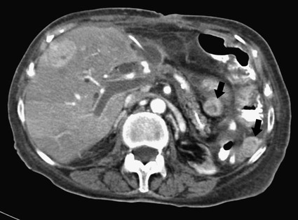
Of the surrounding mesenteric fat arrowheads figure 1 extending along the inferior mesenteric vein open arrow figure 1. Inferior mesenteric vein IMV thrombosis occurs far less often than thrombosis of the superior mesenteric vein 12. However they only show evidence of bowel ischemia in less than 5 of cases. CT and ultrasound have greatly facilitated early diagnosis and the features of superior mesenteric and portal vein thrombosis are well recognized. Thrombosis was con-sidered central when located on the superior mes-enteric vein SMV or inferior mesenteric vein IMV main trunk.
 Source: radiologykey.com
Of the surrounding mesenteric fat arrowheads figure 1 extending along the inferior mesenteric vein open arrow figure 1. Mesenteric vein thrombosis has been described in association with such risk factors as coagulation disorders postoperative dehydration sepsis and trauma. Readers were blinded to clinical information including pathologic conditions. Of the surrounding mesenteric fat arrowheads figure 1 extending along the inferior mesenteric vein open arrow figure 1. Mesenteric vein thrombosis almost always involves the distal small intestine superior mesenteric venous drainage and rarely involves the colon inferior mesenteric venous drainage.
 Source: researchgate.net
The IMV drains blood from the descending colon sigmoid colon and part of the rectum. However they only show evidence of bowel ischemia in less than 5 of cases. Inferior mesenteric vein IMV thrombosis occurs far less often than thrombosis of the superior mesenteric vein 12. It drains the splenic flexure descend-ing colon sigmoid colon and part of the rectum through the left colic sigmoid rectosigmoid and right and left superior rectal veins Fig 1. Late findings include bowel dilataion from ischemia free intraperitoneal air in perforation of an infarcted bowel segment.
 Source: clinicalradiologyonline.net
Inferior mesenteric vein IMV thrombosis occurs far less often than thrombosis of the superior mesenteric vein 12. Imaging of the chest was suggestive of COVID-19 infection which was later confirmed with reverse transcription polymerase chain reaction of his nasopharyngeal swab. MVT accounts for 1 in 5000 to 15000 inpa - tient admissions and 1 in 1000 emer-. In patients with pylephlebitis hypodense zones in the liver might indicate intrahepatic extension of the thrombosis. We present a case of inferior mesenteric vein.
 Source: radiopaedia.org
The anatomic site of involvement in acute mesenteric venous thrombosis is most often ileum 64 to 83 percent or jejunum 50 to 81 percent followed by colon 14 percent and duodenum 4 to 8. CT and ultrasound have greatly facilitated early diagnosis and the features of superior mesenteric and portal vein thrombosis are well recognized. In patients with pylephlebitis hypodense zones in the liver might indicate intrahepatic extension of the thrombosis. Pylephlebitis can be easily detected by color duplex sonography. The anatomic site of involvement in acute mesenteric venous thrombosis is most often ileum 64 to 83 percent or jejunum 50 to 81 percent followed by colon 14 percent and duodenum 4 to 8.
 Source: radiopaedia.org
Radiologists should be familiar with. In patients with pylephlebitis hypodense zones in the liver might indicate intrahepatic extension of the thrombosis. Radiographs are noninvasive and show abnormalities in 50 to 75 of cases. Inferior mesenteric vein thrombosis IMVT is a rare entity that can lead to a potentially lethal event unless recognized early in the disease. Portal venous gas usually.
 Source: radiopaedia.org
However they only show evidence of bowel ischemia in less than 5 of cases. The IMV drains blood from the descending colon sigmoid colon and part of the rectum. Here we present a case of suppurative thrombophlebitis of the inferior mesenteric vein as a complication of sigmoid diverticulitis. It drains the splenic flexure descending colon sigmoid colon and part of the rectum through the left colic sigmoid rectosigmoid and right and left superior rectal veins Fig 1. Although its prevalence is low IMVT presents mainly in certain conditions such as in inflammatory processes like diverticulitis arrhythmias hypercoagulable states connective tissue disorders malignancy or hereditary.
 Source: radiopaedia.org
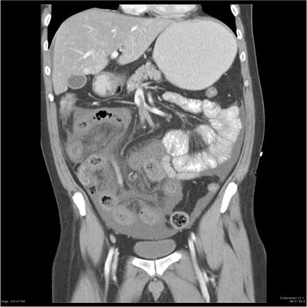
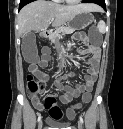
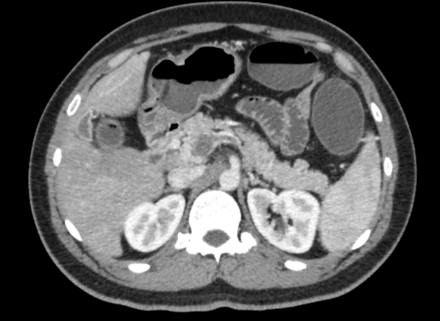
A computed tomography CT scan with intravenous contrast confirmed a thrombus in the inferior mesenteric vein IMV Figure 2 extending to the confluence of. CT and ultrasound have greatly facilitated early diagnosis and the features of superior mesenteric and portal vein thrombosis are well recognized. Mesenteric venous thrombosis MVT describes acute subacute or chronic thrombosis of the superior or inferior mesenteric vein or branches. The anatomic site of involvement in acute mesenteric venous thrombosis is most often ileum 64 to 83 percent or jejunum 50 to 81 percent followed by colon 14 percent and duodenum 4 to 8. The gastrocolic trunk drains into the right-hand aspect of.
 Source: radiopaedia.org
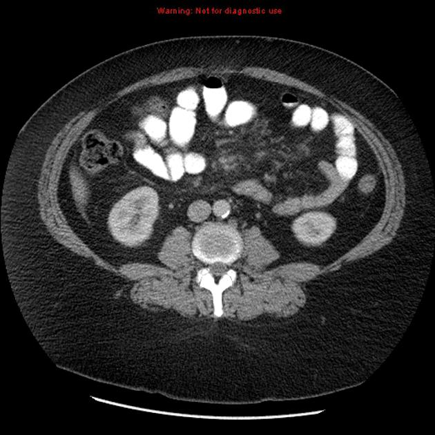
The wall of the inferior mesenteric vein is thickened and the lumen shows filling defects arrowheads figure 2 consistent with thrombosis. The clinical examination was unremarkable but imaging revealed acute mesenteric ischemia caused by superior mesenteric artery and superior mesenteric vein occlusion. Of the surrounding mesenteric fat arrowheads figure 1 extending along the inferior mesenteric vein open arrow figure 1. Early diagnosis with early treatment with anticoagulation or combination with surgery is necessary to prevent bowel infarction. The splenic and portal veins are open and there are no signs of appendicitis.
 Source: researchgate.net
The most informative evaluation for suspected mesenteric venous thrombosis in abdominal imaging. We also review the anatomy of the mesenteric vein. Clinical symptoms of IMV thrombosis are mainly characterized by abdominal pain and the mortality rate remains high 12. Readers were blinded to clinical information including pathologic conditions. Radiographs are noninvasive and show abnormalities in 50 to 75 of cases.
 Source: radiopaedia.org
Inferior mesenteric vein thrombosis IMVT is a rare entity that can lead to a potentially lethal event unless recognized early in the disease. We also review the anatomy of the mesenteric vein. Radiographs are noninvasive and show abnormalities in 50 to 75 of cases. Portal venous gas usually. Clinical symptoms of IMV thrombosis are mainly characterized by abdominal pain and the mortality rate remains high 12.
 Source: radiopaedia.org
Perivascular inflammation can be an indirect sign for pylephlebitis on CT or MRI. Mesenteric venous thrombosis MVT describes acute subacute or chronic thrombosis of the superior or inferior mesenteric vein or branches. Acute mesenteric vein thrombosis is responsible for 6 to 9 of cases of acute mesenteric ischemia1 IMV thrombosis is relatively uncommon and constitutes 4 to 11 of cases of acute mesenteric vein thrombosis23 However IMV thrombosis carries a 15 to 23 risk of mortality46 It is usually seen in the setting of thrombophilia or local inflammation and. The anatomic site of involvement in acute mesenteric venous thrombosis is most often ileum 64 to 83 percent or jejunum 50 to 81 percent followed by colon 14 percent and duodenum 4 to 8. The splenic and portal veins are open and there are no signs of appendicitis.
 Source: radiopaedia.org
We present a case of inferior mesenteric vein. Mesenteric venous thromboses were catego-rized by anatomic location. CT and ultrasound have greatly facilitated early diagnosis and the features of superior mesenteric and portal vein thrombosis are well recognized. Radiologists should be familiar with. Radiographs are noninvasive and show abnormalities in 50 to 75 of cases.
 Source: ejves.com
Nine patients had combination mesenteric vein segment thrombosis. Here we present a case of suppurative thrombophlebitis of the inferior mesenteric vein as a complication of sigmoid diverticulitis. Mesenteric venous thromboses were catego-rized by anatomic location. Readers were blinded to clinical information including pathologic conditions. Thrombolytics were utilized in.
 Source: eventscribe.com
Thrombolytics were utilized in. Here we present a case of suppurative thrombophlebitis of the inferior mesenteric vein as a complication of sigmoid diverticulitis. CT and ultrasound have greatly facilitated early diagnosis and the features of superior mesenteric and portal vein thrombosis are well recognized. Nine patients had combination mesenteric vein segment thrombosis. Mesenteric venous arcades which accompany the arteries unite to form the jejunal and ileal veins in the small bowel mesentery and are joined by the tributaries listed below.
 Source: researchgate.net
Here we present a case of suppurative thrombophlebitis of the inferior mesenteric vein as a complication of sigmoid diverticulitis. Inferior mesenteric vein IMV thrombosis occurs far less often than thrombosis of the superior mesenteric vein 12. The IMV drains blood from the descending colon sigmoid colon and part of the rectum. Acute mesenteric venous thrombosis is uncommon and accounts for 5-10 of cases of acute bowel ischemia 1. Thrombosis was con-sidered central when located on the superior mes-enteric vein SMV or inferior mesenteric vein IMV main trunk.
This site is an open community for users to submit their favorite wallpapers on the internet, all images or pictures in this website are for personal wallpaper use only, it is stricly prohibited to use this wallpaper for commercial purposes, if you are the author and find this image is shared without your permission, please kindly raise a DMCA report to Us.
If you find this site value, please support us by sharing this posts to your favorite social media accounts like Facebook, Instagram and so on or you can also bookmark this blog page with the title inferior mesenteric vein thrombosis radiology by using Ctrl + D for devices a laptop with a Windows operating system or Command + D for laptops with an Apple operating system. If you use a smartphone, you can also use the drawer menu of the browser you are using. Whether it’s a Windows, Mac, iOS or Android operating system, you will still be able to bookmark this website.


