Your Superior mesenteric artery stenosis ultrasound criteria images are ready. Superior mesenteric artery stenosis ultrasound criteria are a topic that is being searched for and liked by netizens today. You can Get the Superior mesenteric artery stenosis ultrasound criteria files here. Find and Download all royalty-free images.
If you’re searching for superior mesenteric artery stenosis ultrasound criteria images information linked to the superior mesenteric artery stenosis ultrasound criteria topic, you have come to the right site. Our website always gives you hints for seeing the maximum quality video and image content, please kindly hunt and find more informative video content and images that fit your interests.
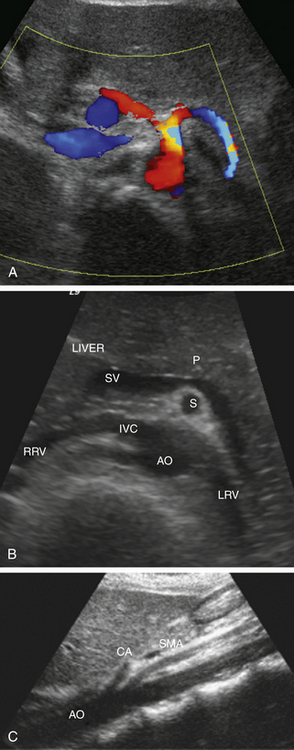
Superior Mesenteric Artery Stenosis Ultrasound Criteria. It has been shown that native vessel criteria overestimate the degree of in-stent restenosis ISR and that velocity criteria for SMA and CA ISR are not well established. There was ultrasound evidence of a proximal superior mesenteric artery SMA stenosis of 70 based on the published criteria. This two-vessel rule holds in most patients and is utilized clinically for the diagnosis of chronic mesenteric ischemia. The gastrointestinal tract is supplied by the celiac trunk the superior mesenteric artery SMA and the inferior mesenteric artery IMA The celiac trunk originates from the anterior aorta just below the diaphragm at the level of the thoracic.
 Linear Endoscopic Ultrasound Eus Findings A Linear Eus Shows The Download Scientific Diagram From researchgate.net
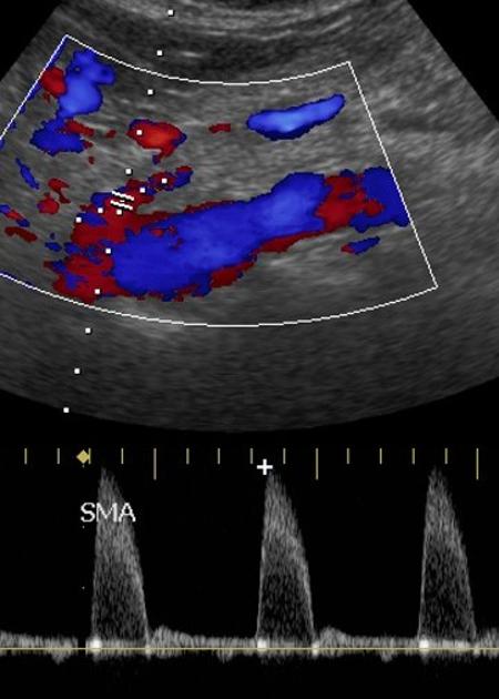
It has been shown that native vessel criteria overestimate the degree of in-stent restenosis ISR and that velocity criteria for SMA and CA ISR are not well established. Fasting duplex criteria for mesenteric stenosis suggest that a superior mesenteric artery peak systolic velocity of 275 cms or greater and a celiac artery peak systolic velocity of 200 cms or greater are reliable indicators of a 70 or greater stenosis9. PSV yielded better results than EDV and MAR. Mesenteric Duplex Ultrasound Celiac artery disease 70 stenosis PSV 200 cmsec Total near total occlusion Absent flow Reversed common hepatic. The source images not shown demonstrated a high-grade right renal artery stenosis but normal left renal and superior mesenteric arteries. However no specific duplex criteria have been developed for detection of mesenteric artery stenosis.
Predicts up to 50 stenosis.
95 and OA 84. Dedicated to the mission of bringing free or low-cost educational materials and information to the global ultrasound community. Criteria for TIPS transjugular intrahepatic portosystemic shunt dysfunction. We aimed to evaluate color-coded Doppler Ultrasonography CDUS for the detection of SMA stenoses and to determine Doppler criteria. The techniques mostly used for the diagnosis of superior mesenteric artery SMA stenosis are computed tomography angiography CTA and magnetic resonance angiography. And for 70 stenosis was 70 cms sens.
 Source: annalsofvascularsurgery.com
95 and OA 84. CDUS is a convenient method with high accuracy for identifying SMA stenosis. 79 and OA 79. The source images not shown demonstrated a high-grade right renal artery stenosis but normal left renal and superior mesenteric arteries. Mesenteric artery duplex scanning appears promising for detection of splanchnic artery stenosis or occlusion or both in patients with symptoms suggestive of chronic intestinal ischemia.
 Source: researchgate.net
RENAL AND MESENTERIC ARTERY STENTS. Mesenteric artery duplex scanning appears promising for detection of splanchnic artery stenosis or occlusion or both in patients with symptoms suggestive of chronic intestinal ischemia. Superior mesenteric artery SMA duplex scanning is utilized to screen for high-grade or70 SMA stenosis peak systolic velocity PSV or275 cmsecond and for follow-up of SMA bypass grafts and stents. PSV yielded better results than EDV and MAR. Moneta et al proposed a SMA peak systolic velocity of 275 cmsec or greater andor no color flow in the SMA as an indicator of 70 percent vessel stenosis.
 Source: researchgate.net
Velocity of 190 cmsec at a stenotic segment or velocity of. CDUS is a convenient method with high accuracy for identifying SMA stenosis. In general severe compromise 70 stenosis or occlusion of at least two of the three mesenteric arteries is required for symptoms of mesenteric ischemia to be present. This pattern usually changes after meals during which the capillary beds are wide open and flow pattern will be noted of low resistance. Velocity.
 Source: thoracickey.com
Duplex ultrasound criteria for diagnosis of splanchnic artery stenosis or occlusion. The optimal threshold values for determining 50-69 SMA stenoses were PSV 280 cms EDV 45 cms and MAR 36. Lack of color Doppler flow. Duplex ultrasound criteria for diagnosis of splanchnic artery stenosis or occlusion. Biri S Biri İ Gultekin Y Yurdakul M Ozdemir M Tola M J Clin Ultrasound 2019 Jun475267-271.
 Source: researchgate.net
The optimal threshold values for determining 50-69 SMA stenoses were PSV 280 cms EDV 45 cms and MAR 36. Doppler ultrasonography criteria of superior mesenteric artery stenosis. The optimal threshold values for determining 50-69 SMA stenoses were PSV 280 cms EDV 45 cms and MAR 36. Your continued use of the site constitutes your acceptance of use of cookies on this site. The distal aorta is perfused by the mesenteric arteries.
 Source: researchgate.net
Velocities were elevated to a maximum of 304 cms with spectral broadening and post-stenotic turbulence. There was ultrasound evidence of a proximal superior mesenteric artery SMA stenosis of 70 based on the published criteria. 95 and OA 84. Velocities were elevated to a maximum of 304 cms with spectral broadening and post-stenotic turbulence. CT angiography CTA confirmed the presence of a.
 Source: radiologykey.com
Duplex ultrasound DUS criteria are well defined for evaluating high-grade stenosis 70 of the native superior mesenteric artery SMA and celiac artery CA. There is however little information correlating duplex scans from stented. The source images not shown demonstrated a high-grade right renal artery stenosis but normal left renal and superior mesenteric arteries. ROC analysis showed that PSV was better than EDV and SMAaortic PSV ratio for 50 stenosis of SMA P 003 and P 0005. The end-diastolic velocities are also elevated in high grade stenosis and a recording of 45 cmsec or greater is suggestive of high degree stenosis Bowersox et al.
 Source: radiopaedia.org
Epub 2019 Jan 29 doi. The end-diastolic velocities are also elevated in high grade stenosis and a recording of 45 cmsec or greater is suggestive of high degree stenosis Bowersox et al. Your continued use of the site constitutes your acceptance of use of cookies on this site. The techniques mostly used for the diagnosis of superior mesenteric artery SMA stenosis are computed tomography angiography CTA and magnetic resonance angiography. Mesenteric Duplex Ultrasound Celiac artery disease 70 stenosis PSV 200 cmsec Total near total occlusion Absent flow Reversed common hepatic.
 Source: researchgate.net
Expected duplex scan findings in SMA bypass grafts have been recently reported. Mesenteric Duplex Ultrasound Celiac artery disease 70 stenosis PSV 200 cmsec Total near total occlusion Absent flow Reversed common hepatic. RENAL AND MESENTERIC ARTERY STENTS. Duplex Criteria for MesentericSplanchnic Arteries. An understanding of mesenteric arterial anatomy is crucial to understanding and managing these patients.
 Source: pinterest.com
Classify lesions in ranges of stenosis severity Atherosclerotic lesions typically at or near the origins of the renal and mesenteric arteries High-grade pressureflow -reducing lesions necessary to produce clinically significant renal and mesenteric ischemia. The distal aorta is perfused by the mesenteric arteries. We aimed to evaluate color-coded Doppler Ultrasonography CDUS for the detection of SMA stenoses and to determine Doppler criteria. It has been shown that native vessel criteria overestimate the degree of in-stent restenosis ISR and that velocity criteria for SMA and CA ISR are not well established. Intestinal brucellosis associated with celiac artery and superior mesenteric artery stenosis and with ileum mucosa.
 Source: researchgate.net
Superior mesenteric artery SMA duplex scanning is utilized to screen for high-grade or70 SMA stenosis peak systolic velocity PSV or275 cmsecond and for follow-up of SMA bypass grafts and stents. Velocity of 190 cmsec at a stenotic segment or velocity of. Fasting duplex criteria for mesenteric stenosis suggest that a superior mesenteric artery peak systolic velocity of 275 cms or greater and a celiac artery peak systolic velocity of 200 cms or greater are reliable indicators of a 70 or greater stenosis9. Mesenteric artery duplex scanning appears promising for detection of splanchnic artery stenosis or occlusion or both in patients with symptoms suggestive of chronic intestinal ischemia. Duplex ultrasound scans have been used for the diagnosis of superior mesenteric artery SMAceliac artery CA stenosis for over 2 decades.
 Source: pinterest.com
Classify lesions in ranges of stenosis severity Atherosclerotic lesions typically at or near the origins of the renal and mesenteric arteries High-grade pressureflow -reducing lesions necessary to produce clinically significant renal and mesenteric ischemia. The gastrointestinal tract is supplied by the celiac trunk the superior mesenteric artery SMA and the inferior mesenteric artery IMA The celiac trunk originates from the anterior aorta just below the diaphragm at the level of the thoracic. Classify lesions in ranges of stenosis severity Atherosclerotic lesions typically at or near the origins of the renal and mesenteric arteries High-grade pressureflow -reducing lesions necessary to produce clinically significant renal and mesenteric ischemia. Predicts up to 50 stenosis. Appropriate threshold velocities for defining various degrees of stenoses have been analyzed leading to the use of specific peak systolic velocities PSV end-diastolic velocities EDV andor CA or SMAaortic systolic ratios.
 Source: pinterest.com
Moneta et al proposed a SMA peak systolic velocity of 275 cmsec or greater andor no color flow in the SMA as an indicator of 70 percent vessel stenosis. CDUS is a convenient method with high accuracy for identifying SMA stenosis. Classify lesions in ranges of stenosis severity Atherosclerotic lesions typically at or near the origins of the renal and mesenteric arteries High-grade pressureflow -reducing lesions necessary to produce clinically significant renal and mesenteric ischemia. The end-diastolic velocities are also elevated in high grade stenosis and a recording of 45 cmsec or greater is suggestive of high degree stenosis Bowersox et al. The EDV threshold that provided the highest OA for detecting 50 stenosis was 45 cms sens.
 Source: pinterest.com
Velocity. Expected duplex scan findings in SMA bypass grafts have been recently reported. Aliasing at the site of the stenosis on color Doppler. Your continued use of the site constitutes your acceptance of use of cookies on this site. And for 70 stenosis was 70 cms sens.
 Source: pinterest.com
Duplex ultrasound scans have been used for the diagnosis of superior mesenteric artery SMAceliac artery CA stenosis for over 2 decades. Your continued use of the site constitutes your acceptance of use of cookies on this site. Classify lesions in ranges of stenosis severity Atherosclerotic lesions typically at or near the origins of the renal and mesenteric arteries High-grade pressureflow -reducing lesions necessary to produce clinically significant renal and mesenteric ischemia. The distal aorta is perfused by the mesenteric arteries. There was ultrasound evidence of a proximal superior mesenteric artery SMA stenosis of 70 based on the published criteria.
 Source: pinterest.com
Appropriate threshold velocities for defining various degrees of stenoses have been analyzed leading to the use of specific peak systolic velocities PSV end-diastolic velocities EDV andor CA or SMAaortic systolic ratios. Duplex ultrasound criteria for diagnosis of splanchnic artery stenosis or occlusion. For identifying 70-99 SMA stenoses they were PSV 395 cms EDV 74 cms and MAR 36. This pattern usually changes after meals during which the capillary beds are wide open and flow pattern will be noted of low resistance. And for 70 stenosis was 70 cms sens.
 Source: pinterest.com
The optimal threshold values for determining 50-69 SMA stenoses were PSV 280 cms EDV 45 cms and MAR 36. We aimed to evaluate color-coded Doppler Ultrasonography CDUS for the detection of SMA stenoses and to determine Doppler criteria. Epub 2019 Jan 29 doi. Velocity. It has been shown that native vessel criteria overestimate the degree of in-stent restenosis ISR and that velocity criteria for SMA and CA ISR are not well established.
 Source: researchgate.net
The source images not shown demonstrated a high-grade right renal artery stenosis but normal left renal and superior mesenteric arteries. An understanding of mesenteric arterial anatomy is crucial to understanding and managing these patients. Epub 2019 Jan 29 doi. Classify lesions in ranges of stenosis severity Atherosclerotic lesions typically at or near the origins of the renal and mesenteric arteries High-grade pressureflow -reducing lesions necessary to produce clinically significant renal and mesenteric ischemia. Velocity.
This site is an open community for users to submit their favorite wallpapers on the internet, all images or pictures in this website are for personal wallpaper use only, it is stricly prohibited to use this wallpaper for commercial purposes, if you are the author and find this image is shared without your permission, please kindly raise a DMCA report to Us.
If you find this site serviceableness, please support us by sharing this posts to your own social media accounts like Facebook, Instagram and so on or you can also bookmark this blog page with the title superior mesenteric artery stenosis ultrasound criteria by using Ctrl + D for devices a laptop with a Windows operating system or Command + D for laptops with an Apple operating system. If you use a smartphone, you can also use the drawer menu of the browser you are using. Whether it’s a Windows, Mac, iOS or Android operating system, you will still be able to bookmark this website.


