Your Superior mesenteric vein first branch images are ready in this website. Superior mesenteric vein first branch are a topic that is being searched for and liked by netizens today. You can Download the Superior mesenteric vein first branch files here. Get all royalty-free vectors.
If you’re searching for superior mesenteric vein first branch pictures information connected with to the superior mesenteric vein first branch keyword, you have pay a visit to the ideal blog. Our site always provides you with hints for viewing the highest quality video and picture content, please kindly search and find more enlightening video content and images that fit your interests.
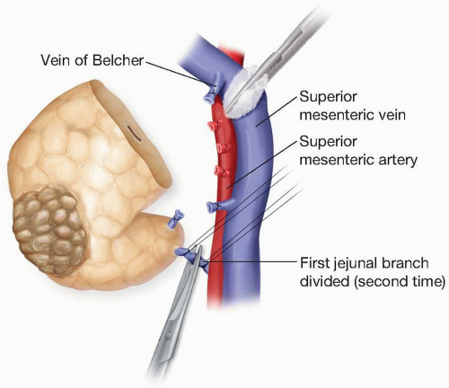
Superior Mesenteric Vein First Branch. The gastrocolic trunk drains into the right-hand aspect of the SMV just anterior to the uncinate process of the pancreas. Courses anterior to the uncinate process of the pancreas and. It unites with the splenic vein posterior to the neck of the pancreas at the level of L1 to form the portal vein. The inferior mesenteric vein.
 Pancreaticobiliary Surgery General Considerations Abdominal Key From abdominalkey.com
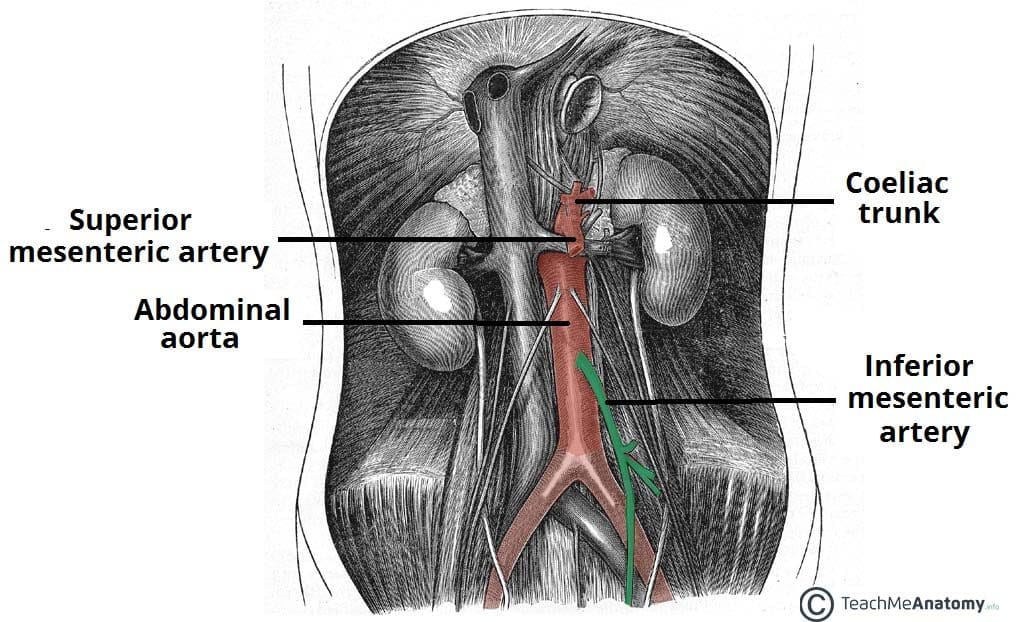
Seven patients had only left or right portal vein thrombosis. This figure was reproduced from 4 with a permission of the publisher. These organs are part of the digestive system. Trace and clean the superior mesenteric artery back to the abdominal aorta. It lies to the right of the superior mesenteric artery. It arises anteriorly from the abdominal aorta at the level of the L1 vertebrae immediately inferior to the origin of the coeliac trunk.
Click card to see definition.
The superior mesenteric vein. Anatomy of the superior mesenteric vein with special reference to the surgical management of first-order branch involvement at pancreaticoduodenectomy. The superior mesenteric vein SMV is a large blood vessel in the abdomen. These organs are part of the digestive system. At the S3 vertebral level the artery divides into two terminal branches one supplying each side of the rectum. The gastrocolic trunk drains into the right-hand aspect of the SMV just anterior to the uncinate process of the pancreas.
 Source: researchgate.net
Each of these arteries give. Often the superior mesenteric vein is considered the common trunk after all the chief tributaries have joined. Anatomy of the Superior Mesenteric Vein With Special Reference to the Surgical Management of First-order Branch Involvement at Pancreaticoduodenectomy. Detailed knowledge of the. Three hundred consecutive contrast-enhanced computed tomography CT scans were reviewed by a surgical oncologist with confirmation of findings by a radiologist.

Obstruction of the portal vein or of its two branches was found in 83 patients 87. The 12 remaining patients had only a single obstructed portal vein branch with or without splenic or superior mesenteric vein obstruction were all symptomatic. Three hundred consecutive contrast-enhanced computed tomography CT scans were reviewed by a surgical oncologist with confirmation of findings by a radiologist. G 439BN 291Gl 1610 This artery is typically the first branch of the superior mesenteric artery. It descends into the pelvis crossing the left common iliac artery and vein.
 Source: researchgate.net
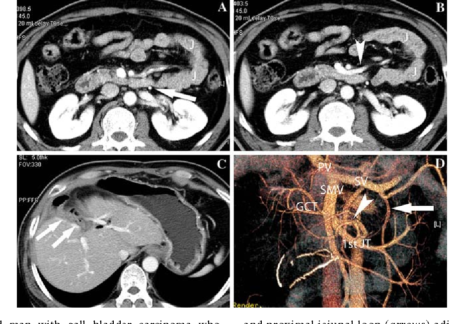
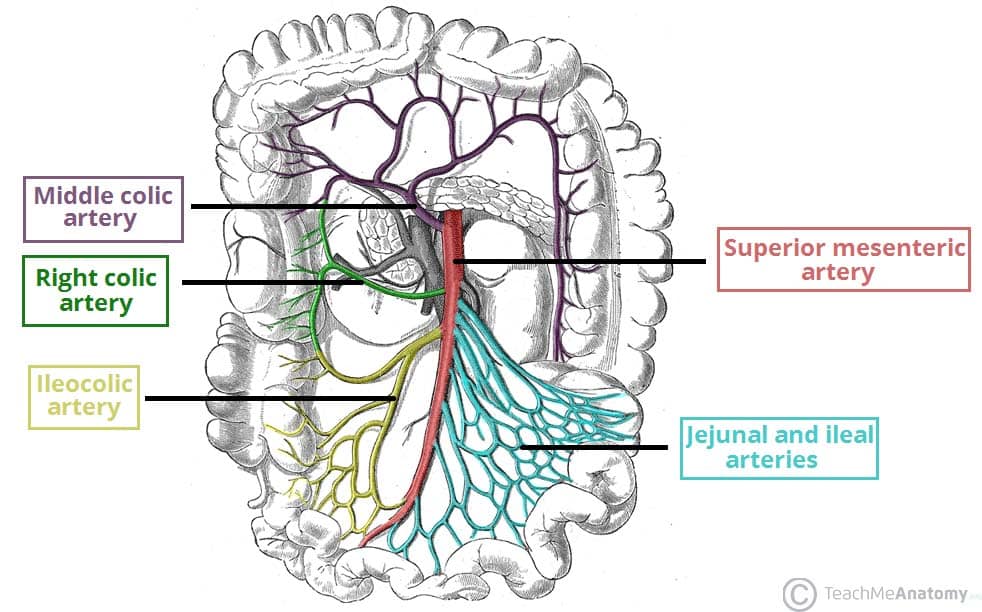
The jejunal and ileal branches of the superior mesenteric artery are variable in number. The gastrocolic trunk drains into the right-hand aspect of the SMV just anterior to the uncinate process of the pancreas. Trace and clean the superior mesenteric artery back to the abdominal aorta. The superior rectal artery is a continuation of the inferior mesenteric artery supplying the rectum. Up to 10 cash back The anatomy of the superior mesenteric vein SMV as observed on cross-sectional CT imaging has been discussed in many studies 35 8 10 11 1315Some of the studies have evaluated the locations of the SMV and the superior mesenteric artery SMA and their relationship with intestinal malrotation 3 8 15The other studies have.
 Source: researchgate.net
The superior mesenteric vein is a large abdominal vein that is formed by the small terminal veins that drain the ileum caecum and vermiform appendix. It unites with the splenic vein posterior to the neck of the pancreas at the level of L1 to form the portal vein. All had clinical symptoms. Blood Supply and Lymphatics. These organs are part of the digestive system.
 Source: slidetodoc.com
At the S3 vertebral level the artery divides into two terminal branches one supplying each side of the rectum. Blood Supply and Lymphatics. Along its course the vein accompanies the superior mesenteric artery that runs on its left side. Up to 10 cash back The anatomy of the superior mesenteric vein SMV as observed on cross-sectional CT imaging has been discussed in many studies 35 8 10 11 1315Some of the studies have evaluated the locations of the SMV and the superior mesenteric artery SMA and their relationship with intestinal malrotation 3 8 15The other studies have. Inferior mesenteric vein C.
 Source: semanticscholar.org
It unites with the splenic vein posterior to the neck of the pancreas at the level of L1 to form the portal vein. The superior mesenteric artery supplies the midgut while the inferior mesenteric artery supplies the hindgut. These organs are part of the digestive system. The superior mesenteric artery provides oxygenated blood and nutrients to the intestines. Artery-First Approaches to Pancreaticoduodenectomy.
 Source: abdominalkey.com
Detailed knowledge of the. The superior rectal artery is a continuation of the inferior mesenteric artery supplying the rectum. The purpose of this study is to determine the anatomic course of the first jejunal branch of the superior mesenteric vein SMV in relation to the superior mesenteric artery SMA. Click card to see definition. The gastrocolic trunk drains into the right-hand aspect of the SMV just anterior to the uncinate process of the pancreas.
 Source: europepmc.org
It arises anteriorly from the abdominal aorta at the level of the L1 vertebrae immediately inferior to the origin of the coeliac trunk. Trace and clean the superior mesenteric artery back to the abdominal aorta. It descends into the pelvis crossing the left common iliac artery and vein. Blood Supply and Lymphatics. The superior mesenteric vein SMV is a large blood vessel in the abdomen.
 Source: researchgate.net
Inferior mesenteric vein C. The artery branches off of the aorta which is the bodys largest blood vessel. Tap card to see definition. The superior mesenteric vein is a large abdominal vein that is formed by the small terminal veins that drain the ileum caecum and vermiform appendix. The superior mesenteric artery arises from the abdominal aorta at the level of the first lumbar vertebral body L1 approximately a centimeter below the coeliac trunk.
 Source: teachmeanatomy.info
It lies to the right of the superior mesenteric artery. Along its course the vein accompanies the superior mesenteric artery that runs on its left side. They pass in the two layers of the mesentery to the jejunum and ileum and progressively divide and join in a series of anastomosing arcades. The 12 remaining patients had only a single obstructed portal vein branch with or without splenic or superior mesenteric vein obstruction were all symptomatic. The superior mesenteric vein is a large abdominal vein that is formed by the small terminal veins that drain the ileum caecum and vermiform appendix.
 Source: kenhub.com
Right common carotid artery. It arises anteriorly from the abdominal aorta at the level of the L1 vertebrae immediately inferior to the origin of the coeliac trunk. This artery bifurcates into anterior and posterior branches both of which form anastomoses with branches of the superior pancreaticoduodenal artery a branch of the celiac artery. It arises above the renal arteries that arise at vertebral level L1-L2. The purpose of this study is to determine the anatomic course of the first jejunal branch of the superior mesenteric vein SMV in relation to the superior mesenteric artery SMA.
 Source: pancreas.imedpub.com
At the S3 vertebral level the artery divides into two terminal branches one supplying each side of the rectum. Hepatic portal system. Often the superior mesenteric vein is considered the common trunk after all the chief tributaries have joined. The first branch of the superior mesenteric artery is the inferior pancreaticoduodenal artery. Tap card to see definition.
 Source: researchgate.net
The 12 remaining patients had only a single obstructed portal vein branch with or without splenic or superior mesenteric vein obstruction were all symptomatic. Along its course the vein accompanies the superior mesenteric artery that runs on its left side. Blood Supply and Lymphatics. Click again to see term. This figure was reproduced from 4 with a permission of the publisher.
 Source: slidetodoc.com
The superior rectal artery is a continuation of the inferior mesenteric artery supplying the rectum. All had clinical symptoms. G 439BN 291Gl 1610 This artery is typically the first branch of the superior mesenteric artery. Hepatic portal system. Each of these arteries give.

It unites with the splenic vein posterior to the neck of the pancreas at the level of L1 to form the portal vein. The inferior mesenteric vein. It descends into the pelvis crossing the left common iliac artery and vein. The superior rectal artery is a continuation of the inferior mesenteric artery supplying the rectum. Along its course the vein accompanies the superior mesenteric artery that runs on its left side.
 Source: teachmeanatomy.info
Segmental resection of one of the 2 first-order branches of the SMV may be performed without venous reconstruction if mesenteric venous flow is preserved through the remaining first-order branch. The superior mesenteric artery supplies the midgut while the inferior mesenteric artery supplies the hindgut. S superior approach A anterior approach P posterior approach L left posterior approach R rightmedial uncinate approach M mesenteric approach. The 12 remaining patients had only a single obstructed portal vein branch with or without splenic or superior mesenteric vein obstruction were all symptomatic. It descends into the pelvis crossing the left common iliac artery and vein.
 Source: slidetodoc.com
Superior Rectal Artery. Inferior mesenteric vein C. Detailed knowledge of the. The first vessel to branch from the aortic arch is the A. The artery branches off of the aorta which is the bodys largest blood vessel.
 Source: researchgate.net
It descends into the pelvis crossing the left common iliac artery and vein. Artery-First Approaches to Pancreaticoduodenectomy. The superior mesenteric vein SMV is a large blood vessel in the abdomen. Three hundred consecutive contrast-enhanced computed tomography CT scans were reviewed by a surgical oncologist with confirmation of findings by a radiologist. All had clinical symptoms.
This site is an open community for users to share their favorite wallpapers on the internet, all images or pictures in this website are for personal wallpaper use only, it is stricly prohibited to use this wallpaper for commercial purposes, if you are the author and find this image is shared without your permission, please kindly raise a DMCA report to Us.
If you find this site value, please support us by sharing this posts to your preference social media accounts like Facebook, Instagram and so on or you can also save this blog page with the title superior mesenteric vein first branch by using Ctrl + D for devices a laptop with a Windows operating system or Command + D for laptops with an Apple operating system. If you use a smartphone, you can also use the drawer menu of the browser you are using. Whether it’s a Windows, Mac, iOS or Android operating system, you will still be able to bookmark this website.


