Your Superior mesenteric vein thrombosis ct radiology images are available. Superior mesenteric vein thrombosis ct radiology are a topic that is being searched for and liked by netizens now. You can Get the Superior mesenteric vein thrombosis ct radiology files here. Get all free vectors.
If you’re searching for superior mesenteric vein thrombosis ct radiology pictures information connected with to the superior mesenteric vein thrombosis ct radiology topic, you have pay a visit to the ideal blog. Our website always gives you hints for downloading the maximum quality video and image content, please kindly hunt and find more enlightening video content and images that fit your interests.
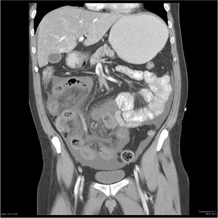
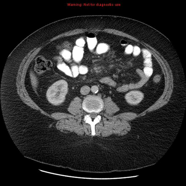
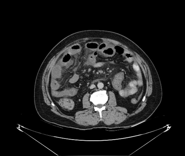
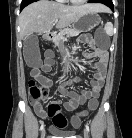
Superior Mesenteric Vein Thrombosis Ct Radiology. Further investigation ruled out haematological causes and COVID-19 was determined to be the cause. A vascular medicine specialist was consulted. Further investigation ruled out haematological causes and COVID-19 was. Axial CT confirmed a thrombus within the superior mesenteric vein.
 Veno Occlusive Mesenteric Ischemia Radiology Reference Article Radiopaedia Org From radiopaedia.org
Abdominal contrast-enhanced CT was performed. Axial contrast-enhanced CT images show a hypodense thrombus within the superior mesenteric vein SMV Fig. When you have mesenteric venous thrombosis MVT you have a blood clot in a vein around where your intestines attach to your belly. Axial CT confirmed a thrombus within the superior mesenteric vein. MVT may present with acute abdominal pain. Further investigation ruled out haematological causes and COVID-19 was.
SAGAR Radiology Department City Hospital NHS Trust Birmingham UK Thrombosis in Acute We describe the computed tomography CT appearances of four patients with acute or acute on chronic case 3 pancreatitis which.
Each of the aforementioned conditions re-quires a different approach to diagnosis and management. In three of the four cases follow-up CT scans showed the SMV thrombosis to. SAGAR Radiology Department City Hospital NHS Trust Birmingham UK Thrombosis in Acute We describe the computed tomography CT appearances of four patients with acute or acute on chronic case 3 pancreatitis which. The bowel was dilated with mural thickening and with some gas in the bowel wall. MVT may present with acute abdominal pain. He had contracted COVID-19 9 days prior.
 Source: researchgate.net
A CT scan of the abdomen with coronal view showed superior mesenteric vein thrombosis arrow. Superior mesenteric venous SMV thrombosis is a rare but potentially deadly complication of laparoscopic bariatric surgery. Chronic mesenteric ischemia also known as intestinal angina is most often due to arterial atherosclerotic disease. Department of Radiology Royal Perth Hospital Perth Western Australia Australia. The most informative evaluation for suspected mesenteric venous thrombosis in abdominal imaging.
 Source: researchgate.net
Often the superior mesenteric vein is considered the common trunk after all the chief tributaries have joined. It is a rare condition. Mesenteric venous arcades which accompany the arteries unite to form the jejunal and ileal veins in the small bowel mesentery and are joined by the tributaries listed below. Reversible superior mesenteric vein thrombosis in acute pancreatitis. Because many patients present post-surgery with abdominal pain radiologists need to be knowledgeable of the imaging findings on CT of SMV thrombosis so they dont overlook this complication.
 Source: radiopaedia.org
Overview Mesenteric venous thrombosis MVT describes acute subacute or chronic thrombosis of the superior or inferior mesenteric vein or branches. A 68-year-old man was referred to the general surgeons on account of his abdominal pain of unknown cause. CT chest abdomen and pelvis revealed an extensive thrombus extending from the portal vein to the superior mesenteric vein. There is hypoattenuation of the ileum and ascending colon with mesenteric stranding and ascites consistent with intestinal ischemia. SAGAR Radiology Department City Hospital NHS Trust Birmingham UK Thrombosis in Acute We describe the computed tomography CT appearances of four patients with acute or acute on chronic case 3 pancreatitis which.
 Source: radiopaedia.org
Each of the aforementioned conditions re-quires a different approach to diagnosis and management. VMI was diagnosed in the presence of SMV. SAGAR Radiology Department City Hospital NHS Trust Birmingham UK Thrombosis in Acute We describe the computed tomography CT appearances of four patients with acute or acute on chronic case 3 pancreatitis which. He had contracted COVID-19 9 days prior. Isolated superior mesenteric venous thrombosis SMVT is when a blood clot forms in the SMV.
 Source: angiologist.com
3 arrow and the portal vein Fig. SAGAR Radiology Department City Hospital NHS Trust Birmingham UK Thrombosis in Acute We describe the computed tomography CT appearances of four patients with acute or acute on chronic case 3 pancreatitis which. Various causes of mesenteric vein thrombosis are classified underlying pathogenic mechanisms are enumerated and discussed and multidetector CT features of venous bowel ischemia are described and. Abdominal contrast-enhanced CT was performed. It is a rare condition.

Although no correlation was noted between risk factor and outcome the presence of bowel wall thickening and mesenteric congestion on CT or MR imaging was as-. The bowel was dilated with mural thickening and with some gas in the bowel wall. Overview Mesenteric venous thrombosis MVT describes acute subacute or chronic thrombosis of the superior or inferior mesenteric vein or branches. Although no correlation was noted between risk factor and outcome the presence of bowel wall thickening and mesenteric congestion on CT or MR imaging was as-. Various causes of mesenteric vein thrombosis are classified underlying pathogenic mechanisms are enumerated and discussed and multidetector CT features of venous bowel ischemia are described and.
 Source: researchgate.net
When you have mesenteric venous thrombosis MVT you have a blood clot in a vein around where your intestines attach to your belly. A vascular medicine specialist was consulted. Mesenteric vein thrombosis MVT accounts for 515 of all mesenteric ischemic events and is classified as either primary or secondary. A 68-year-old man was referred to the general surgeons on account of his abdominal pain of unknown cause. Because many patients present post-surgery with abdominal pain radiologists need to be knowledgeable of the imaging findings on CT of SMV thrombosis so they dont overlook this complication.
 Source: radiopaedia.org
A 68-year-old man was referred to the general surgeons on account of his abdominal pain of unknown cause. Gross anatomy Origin and course. CT chest abdomen and pelvis revealed an extensive thrombus extending from the portal vein to the superior mesenteric vein. Further investigation ruled out haematological causes and COVID-19 was determined to be the cause. Radiographs are noninvasive and show abnormalities in 50 to 75 of cases.
 Source: radiopaedia.org
The most informative evaluation for suspected mesenteric venous thrombosis in abdominal imaging. Because many patients present post-surgery with abdominal pain radiologists need to be knowledgeable of the imaging findings on CT of SMV thrombosis so they dont overlook this complication. It is a rare condition. The most common predisposing factors in superior mesenteric vein thrombosis with radiologically occult cause are recent abdominal surgery infection and hy-percoagulable states. During the course of her hospitalization she was seen by a vascular surgeon gastroenterologist and hematologist.
 Source: radiopaedia.org
He had contracted COVID-19 9 days prior. A vascular medicine specialist was consulted. There was no evidence of air in or adjacent to the thrombus and no evidence of liver abscess. Mesenteric vein thrombosis MVT accounts for 515 of all mesenteric ischemic events and is classified as either primary or secondary. Mesenteric venous arcades which accompany the arteries unite to form the jejunal and ileal veins in the small bowel mesentery and are joined by the tributaries listed below.
 Source: ejves.com
Department of Radiology Royal Perth Hospital Perth Western Australia Australia. MVT may present with acute abdominal pain. Reversible superior mesenteric vein thrombosis in acute pancreatitis. Each of the aforementioned conditions re-quires a different approach to diagnosis and management. He had contracted COVID-19 9 days prior.
 Source: ejves.com
There is hypoattenuation of the ileum and ascending colon with mesenteric stranding and ascites consistent with intestinal ischemia. No abnormality was detected in the right iliac fossa. SMVT can occur as a result of cancer peritonitis increased blood clotting hypercoagulable state protein C deficiency polycythemia vera recent abdominal surgery high blood pressure in the portal vein portal. Often the superior mesenteric vein is considered the common trunk after all the chief tributaries have joined. Thrombus was seen in the superior mesenteric artery and renal vessels and infarction was noted in the kidneys.
 Source: researchgate.net
When you have mesenteric venous thrombosis MVT you have a blood clot in a vein around where your intestines attach to your belly. Each of the aforementioned conditions re-quires a different approach to diagnosis and management. Axial contrast-enhanced CT images show a hypodense thrombus within the superior mesenteric vein SMV Fig. It is a rare condition. Axial CT confirmed a thrombus within the superior mesenteric vein.
 Source: radiopaedia.org
Gross anatomy Origin and course. Although no correlation was noted between risk factor and outcome the presence of bowel wall thickening and mesenteric congestion on CT or MR imaging was as-. MVT may present with acute abdominal pain. Abdominal contrast-enhanced CT was performed. The present case report describes the unusual progression of COVID-19 disease from pneumonia to a procoagulant state leading to superior mesenteric artery thrombosis and subsequent gut.
 Source: radiopaedia.org
Although no correlation was noted between risk factor and outcome the presence of bowel wall thickening and mesenteric congestion on CT or MR imaging was as-. The bowel was dilated with mural thickening and with some gas in the bowel wall. A vascular medicine specialist was consulted. Reversible superior mesenteric vein thrombosis in acute pancreatitis. Axial CT confirmed a thrombus within the superior mesenteric vein.
 Source: radiopaedia.org
Each of the aforementioned conditions re-quires a different approach to diagnosis and management. The most informative evaluation for suspected mesenteric venous thrombosis in abdominal imaging. Clinical Radiology 1995 50 628-633 Reversible Superior Mesenteric Vein Pancreatitis. Superior mesenteric venous SMV thrombosis is a rare but potentially deadly complication of laparoscopic bariatric surgery. The patient was then placed on a heparin drip.
 Source: radiopaedia.org
No abnormality was detected in the right iliac fossa. The CT Appearances P. The most common predisposing factors in superior mesenteric vein thrombosis with radiologically occult cause are recent abdominal surgery infection and hy-percoagulable states. 3 arrow and the portal vein Fig. The patient was then placed on a heparin drip.

Axial CT confirmed a thrombus within the superior mesenteric vein. A CT scan of the abdomen showed superior mesenteric vein thrombosis arrow. Various causes of mesenteric vein thrombosis are classified underlying pathogenic mechanisms are enumerated and discussed and multidetector CT features of venous bowel ischemia are described and. The bowel was dilated with mural thickening and with some gas in the bowel wall. The patient was then placed on a heparin drip.
This site is an open community for users to share their favorite wallpapers on the internet, all images or pictures in this website are for personal wallpaper use only, it is stricly prohibited to use this wallpaper for commercial purposes, if you are the author and find this image is shared without your permission, please kindly raise a DMCA report to Us.
If you find this site good, please support us by sharing this posts to your favorite social media accounts like Facebook, Instagram and so on or you can also bookmark this blog page with the title superior mesenteric vein thrombosis ct radiology by using Ctrl + D for devices a laptop with a Windows operating system or Command + D for laptops with an Apple operating system. If you use a smartphone, you can also use the drawer menu of the browser you are using. Whether it’s a Windows, Mac, iOS or Android operating system, you will still be able to bookmark this website.


