Your Superior mesenteric vein thrombosis radiopaedia images are ready. Superior mesenteric vein thrombosis radiopaedia are a topic that is being searched for and liked by netizens today. You can Download the Superior mesenteric vein thrombosis radiopaedia files here. Find and Download all free photos.
If you’re searching for superior mesenteric vein thrombosis radiopaedia pictures information linked to the superior mesenteric vein thrombosis radiopaedia topic, you have come to the ideal blog. Our website always provides you with suggestions for viewing the highest quality video and image content, please kindly surf and find more enlightening video articles and graphics that match your interests.
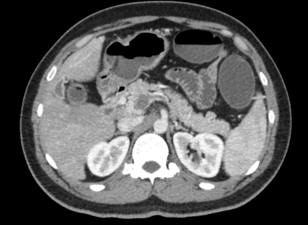
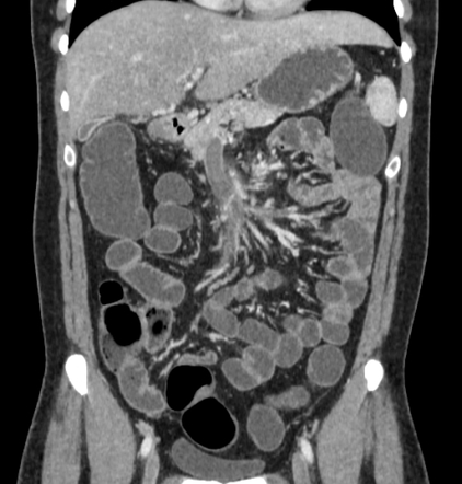
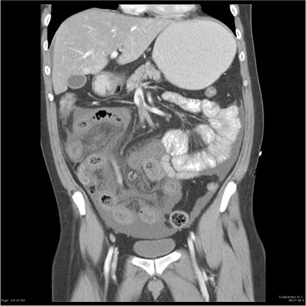
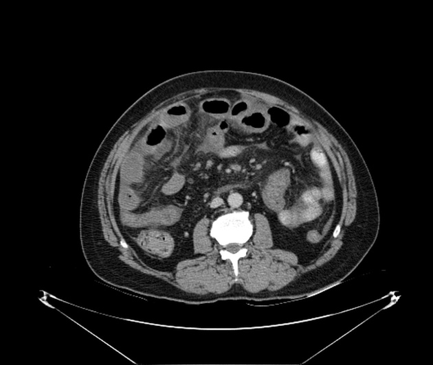
Superior Mesenteric Vein Thrombosis Radiopaedia. The hallmark is pain out. The left hepatic portal splenic and superior mesenteric veins contain large volume thrombus. The superior mesenteric vein SMV accompanies the superior mesenteric artery SMA and drains the midgut to the portal venous system. Variable attenuation of the liver due to the venous thrombus and thick-walled jejunum in the left side of the abdomen likely due to venous congestion secondary to.
 Superior Mesenteric Vein Thrombosis Radiology Case Radiopaedia Org From radiopaedia.org
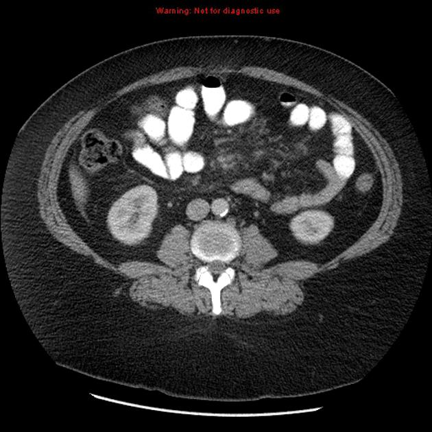
The ovarian vein is best visualized at level of the origin of the inferior mesenteric artery where it is surrounded by retroperitoneal fat and in the pelvis medial to external iliac vessels 25. Ncbinlmnihgov While mesenteric arterial thrombosis results from arrhythmia and cardiac etiologies mesenteric venous thrombosis is overwhelmingly. Non opacification of main trunk of superior mesenteric vein up to the portosplenic confluence with perivascular fat stranding. Acute superior mesenteric vein thrombosis is one of the less common causes of acute mesenteric ischemia. SMV thrombosis accounts for approximately 10 of all mesenteric ischemia. The splenic vein is dilated 103mm.
Superior mesenteric vein Vena mesenterica superior The superior mesenteric vein SMV is a large venous vessel located in the abdomenIt arises within the mesentery of the small intestine from the small tributaries that drain blood from the terminal ileum caecum and vermiform appendixIt terminates by uniting with the splenic vein and forming the portal vein.
The portal vein has large central thrombus with a rim of surrounding luminal contrast in keeping with the Polo Mint sign. Spleen is enlarged spanning 20cm. Possible causes include Acute Pancreatitis. Mesenteric venous thrombosis is a rare occurrence that can cause a variety of symptoms including progressively worsening diffuse colicky abdominal pain. The hallmark is pain out. Superior mesenteric vein Vena mesenterica superior The superior mesenteric vein SMV is a large venous vessel located in the abdomenIt arises within the mesentery of the small intestine from the small tributaries that drain blood from the terminal ileum caecum and vermiform appendixIt terminates by uniting with the splenic vein and forming the portal vein.
 Source: ejves.com
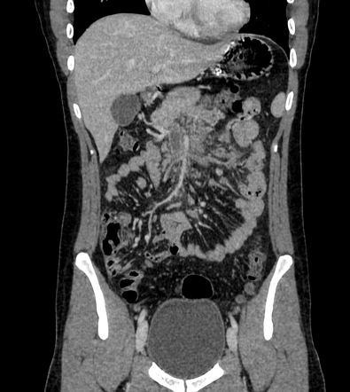
Despite thrombosis of the SMV small bowel necrosis is less likely to occur presumably due to persistent arterial supply and some venous drainage via collaterals. SMV thrombosis accounts for approximately 10 of all mesenteric ischemia. This article is focused on acute mesenteric ischemia. The intervention used was infrainguinal composite artery-vein bypass grafting to popliteal n 18 and infrapopliteal n 30 arteries with an occluded segment of the superficial femoral artery prepared with eversion endarterectomy and an autogenous vein conduit harvested from greater saphenous veins n 43 arm veins n 3 and lesser. There is evidence of superior mesenteric vein and portal vein thrombosis causing proximal small bowel dilatation and wall thickening with poor enhancement of dilated small bowel loops with suspicious pneumatosis intestinalis highly suggestive for ischemia.
 Source: radiopaedia.org
Non opacification of main trunk of superior mesenteric vein up to the portosplenic confluence with perivascular fat stranding. Despite thrombosis of the SMV small bowel necrosis is less likely to occur presumably due to persistent arterial supply and some venous drainage via collaterals. Related pathology ovarian vein phlebolith ovarian vein reflux and pelvic congestion syndrome. Spleen is enlarged spanning 20cm. Ated with portal or mesenteric venous thrombosis Spontaneous idiopathic thrombosis of the such as pancreatitis hypercoagulability states cirrhosis or surgery are said to have secondary.
 Source: radiopaedia.org
Main portal vein is dilated 145mm. Mesenteric venous thrombosis is a rare occurrence that can cause a variety of symptoms including progressively worsening diffuse colicky abdominal pain. Mesenteric Vein Thrombosis Symptom Checker. CT shows wall thickening of a 15cm segment of small bowel centrally within the abdomen. Despite thrombosis of the SMV small bowel necrosis is less likely to occur presumably due to persistent arterial supply and some venous drainage via collaterals.
 Source: angiologist.com
Mesenteric Vein Thrombosis Symptom Checker. Although the mainstay for treating patients with mesenteric venous thrombosis has been surgical resection of affected bowel technical innovations have added. CT shows wall thickening of a 15cm segment of small bowel centrally within the abdomen. Variable attenuation of the liver due to the venous thrombus and thick-walled jejunum in the left side of the abdomen likely due to venous congestion secondary to. There is evidence of superior mesenteric vein and portal vein thrombosis causing proximal small bowel dilatation and wall thickening with poor enhancement of dilated small bowel loops with suspicious pneumatosis intestinalis highly suggestive for ischemia.
 Source: researchgate.net
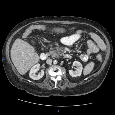
Non-enhancement of the superior mesenteric vein but enhancement of the splenic and portal veins and mild mesenteric fat stranding adjacent to the SMV. Acute superior mesenteric vein thrombosis is one of the less common causes of acute mesenteric ischemia. The ovarian vein is best visualized at level of the origin of the inferior mesenteric artery where it is surrounded by retroperitoneal fat and in the pelvis medial to external iliac vessels 25. This article is focused on acute mesenteric ischemia. Rarely the superior mesenteric artery presses against a renal vein or the duodenum causing potentially life-threatening problems.
 Source: radiopaedia.org
Despite thrombosis of the SMV small bowel necrosis is less likely to occur presumably due to persistent arterial supply and some venous drainage via collaterals. Related pathology ovarian vein phlebolith ovarian vein reflux and pelvic congestion syndrome. Ncbinlmnihgov While mesenteric arterial thrombosis results from arrhythmia and cardiac etiologies mesenteric venous thrombosis is overwhelmingly. When you have mesenteric venous thrombosis MVT you have a blood clot in a vein around where your intestines attach to your belly. The superior mesenteric vein SMV accompanies the superior mesenteric artery SMA and drains the midgut to the portal venous system.
 Source: radiopaedia.org
It usually presents as nausea vomiting and vague abdominal pain that can easily be masked by pancreatitis as in our patient. The testicular vein normally measures 1-3 mm in diameter 8. Possible causes include Acute Pancreatitis. There is evidence of superior mesenteric vein and portal vein thrombosis causing proximal small bowel dilatation and wall thickening with poor enhancement of dilated small bowel loops with suspicious pneumatosis intestinalis highly suggestive for ischemia. This article is focused on acute mesenteric ischemia.
 Source: researchgate.net
The superior mesenteric vein is dilated and non-enhancing in keeping with SMV thrombosis complicated by a segment of small bowel ischemia. It usually presents as nausea vomiting and vague abdominal pain that can easily be masked by pancreatitis as in our patient. SMV thrombosis accounts for approximately 10 of all mesenteric ischemia. The superior mesenteric vein SMV accompanies the superior mesenteric artery SMA and drains the midgut to the portal venous system. Variable attenuation of the liver due to the venous thrombus and thick-walled jejunum in the left side of the abdomen likely due to venous congestion secondary to.
 Source: radiopaedia.org
This article is focused on acute mesenteric ischemia. SMV thrombosis accounts for approximately 10 of all mesenteric ischemia. Despite thrombosis of the SMV small bowel necrosis is less likely to occur presumably due to persistent arterial supply and some venous drainage via collaterals. Related pathology ovarian vein phlebolith ovarian vein reflux and pelvic congestion syndrome. Mesenteric venous thrombosis can be classified with known medical conditions or factors associ on the basis of its cause as primary or secondary.
 Source: researchgate.net
Main portal vein is dilated 145mm. Acute superior mesenteric vein thrombosis presents vaguely as an acute abdomen with gradually worsening diffuse colicky abdominal pain associated with distention and symptoms may have been present for a few days 23. There is evidence of superior mesenteric vein and portal vein thrombosis causing proximal small bowel dilatation and wall thickening with poor enhancement of dilated small bowel loops with suspicious pneumatosis intestinalis highly suggestive for ischemia. Variable attenuation of the liver due to the venous thrombus and thick-walled jejunum in the left side of the abdomen likely due to venous congestion secondary to. Minimal free fluid in around the liver and in the right pericolic gutter.
 Source: radiopaedia.org
It usually presents as nausea vomiting and vague abdominal pain that can easily be masked by pancreatitis as in our patient. The hallmark is pain out. The superior mesenteric vein SMV accompanies the superior mesenteric artery SMA and drains the midgut to the portal venous system. Non opacification of main trunk of superior mesenteric vein up to the portosplenic confluence with perivascular fat stranding. The testicular vein normally measures 1-3 mm in diameter 8.
 Source: radiopaedia.org
Although the mainstay for treating patients with mesenteric venous thrombosis has been surgical resection of affected bowel technical innovations have added. Main portal vein is dilated 145mm. This article is focused on acute mesenteric ischemia. Variable attenuation of the liver due to the venous thrombus and thick-walled jejunum in the left side of the abdomen likely due to venous congestion secondary to. Possible causes include Acute Pancreatitis.
 Source: radiopaedia.org
Possible causes include Acute Pancreatitis. Acute superior mesenteric vein thrombosis is one of the less common causes of acute mesenteric ischemia. Variable attenuation of the liver due to the venous thrombus and thick-walled jejunum in the left side of the abdomen likely due to venous congestion secondary to. Mesenteric venous thrombosis is a rare occurrence that can cause a variety of symptoms including progressively worsening diffuse colicky abdominal pain. Transient hepatic attenuation differences THAD are seen within the liver which is likely secondary to reduced portal vein flow.
 Source: radiopaedia.org
Our purpose was to examine the clinical presentation imaging appearance etiology and clinical outcome in patients who had acute thrombosis of the superior mesenteric vein with radiologically occult cause. Mesenteric ischemia also commonly referred to as bowel or intestinal ischemia refers to vascular compromise of the bowel and its mesentery that in the acute setting has a very high mortality if not treated expedientlyMesenteric ischemia is far more commonly acute than chronic in etiology. Main portal vein is dilated 145mm. Non opacification of main trunk of superior mesenteric vein up to the portosplenic confluence with perivascular fat stranding. Acute superior mesenteric vein thrombosis presents vaguely as an acute abdomen with gradually worsening diffuse colicky abdominal pain associated with distention and symptoms may have been present for a few days 23.
 Source: radiopaedia.org
Mesenteric Vein Thrombosis Symptom Checker. The superior mesenteric artery plays a vital role in keeping the digestive system healthy and functioning. Non-enhancement of the superior mesenteric vein but enhancement of the splenic and portal veins and mild mesenteric fat stranding adjacent to the SMV. When you have mesenteric venous thrombosis MVT you have a blood clot in a vein around where your intestines attach to your belly. The left hepatic portal splenic and superior mesenteric veins contain large volume thrombus.
 Source: radiopaedia.org
The portal vein has large central thrombus with a rim of surrounding luminal contrast in keeping with the Polo Mint sign. Mesenteric ischemia also commonly referred to as bowel or intestinal ischemia refers to vascular compromise of the bowel and its mesentery that in the acute setting has a very high mortality if not treated expedientlyMesenteric ischemia is far more commonly acute than chronic in etiology. Mesenteric venous thrombosis is a rare occurrence that can cause a variety of symptoms including progressively worsening diffuse colicky abdominal pain. The portal vein has large central thrombus with a rim of surrounding luminal contrast in keeping with the Polo Mint sign. The testicular vein normally measures 1-3 mm in diameter 8.

The ovarian vein is best visualized at level of the origin of the inferior mesenteric artery where it is surrounded by retroperitoneal fat and in the pelvis medial to external iliac vessels 25. It usually presents as nausea vomiting and vague abdominal pain that can easily be masked by pancreatitis as in our patient. The SMV thrombosis diagnosis is difficult due to the lack of specificity of clinical symptoms. The superior mesenteric vein thrombosis is a well know complication of laparoscopic procedures though a rare cause of intestinal ischemia. Its branches are patent.
 Source: radiopaedia.org
The intervention used was infrainguinal composite artery-vein bypass grafting to popliteal n 18 and infrapopliteal n 30 arteries with an occluded segment of the superficial femoral artery prepared with eversion endarterectomy and an autogenous vein conduit harvested from greater saphenous veins n 43 arm veins n 3 and lesser. The testicular vein normally measures 1-3 mm in diameter 8. The SMV thrombosis diagnosis is difficult due to the lack of specificity of clinical symptoms. The superior mesenteric artery plays a vital role in keeping the digestive system healthy and functioning. If the artery clogs with plaque or develops a clot blood flow to digestive organs slows.
This site is an open community for users to do sharing their favorite wallpapers on the internet, all images or pictures in this website are for personal wallpaper use only, it is stricly prohibited to use this wallpaper for commercial purposes, if you are the author and find this image is shared without your permission, please kindly raise a DMCA report to Us.
If you find this site adventageous, please support us by sharing this posts to your favorite social media accounts like Facebook, Instagram and so on or you can also save this blog page with the title superior mesenteric vein thrombosis radiopaedia by using Ctrl + D for devices a laptop with a Windows operating system or Command + D for laptops with an Apple operating system. If you use a smartphone, you can also use the drawer menu of the browser you are using. Whether it’s a Windows, Mac, iOS or Android operating system, you will still be able to bookmark this website.


