Your What is a photoreceptor layer images are available in this site. What is a photoreceptor layer are a topic that is being searched for and liked by netizens today. You can Get the What is a photoreceptor layer files here. Get all royalty-free photos and vectors.
If you’re searching for what is a photoreceptor layer images information related to the what is a photoreceptor layer keyword, you have visit the ideal site. Our website always provides you with hints for seeing the highest quality video and picture content, please kindly hunt and find more informative video articles and images that match your interests.
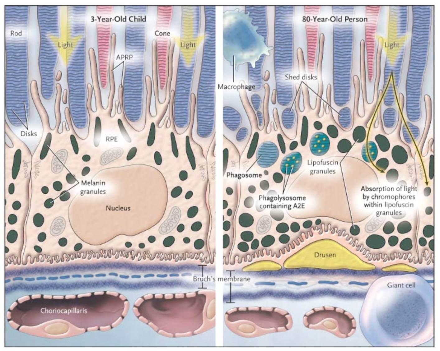
What Is A Photoreceptor Layer. One isomer 11-cis-retinal combines with opsin in the retinal rods scotopsin to form rhodopsin visual purple. Photoreceptors are specialized cells for detecting light. The photoreceptor consists of 1 an outer segment filled with stacks of membranes like a stack of poker chips containing the visual pigment molecules such as rhodopsins 2 an inner segment containing mitochondria ribosomes and membranes where opsin molecules are assembled and passed to be part of the outer segment discs 3 a cell body containing the nucleus of the photoreceptor cell and 4 a. 7 rows Photoreceptor layer of retina - histological slide.
 Pin De Keryee Morton Em Unit 4 Anatomy From pinterest.com
Photoreception is the process that describes how photoreceptors like rods and cones absorb light waves that enter the eye and convert them into electrical signals which are then sent to the brain for visual processing. A photoreceptor cell is a specialized type of neuroepithelial cell found in the retina that is capable of visual phototransduction. There are currently three known types. 1 The pigmented epithelium which is adjacent to the choroid absorbs light to reduce back reflection of light onto the retina 2 the photoreceptor layer contains photosensitive outer segments of rods and cones 3 the outer nuclear layer contains cell bodies of. 7 rows Photoreceptor layer of retina - histological slide. First of all there are two types of receptors.
To describe changes of the foveal photoreceptor layer using optical coherence tomography OCT in central serous chorioretinopathy CSC and evaluate the correlation with visual acuity VA loss.
Photoreceptors are special cells in the eyes retina that are responsible for converting light into signals that are sent to the brain. The cellular layers of the retina are as follows. Pertaining to the retina. The cell bodies of the two latter neuronal cells as well as from amacrine and Müller cells constitute the inner nuclear layer. There are currently three known types. Photoreceptors Layer Layer of rods and cones.
 Source: pinterest.com
Pertaining to the retina. Retinal pigment epithelium This is a single layer of cells that provide essential nutrition and waste removal for the photoreceptor cells. Another all-trans-retinal or visual yellow results from the bleaching of rhodopsin by light in which the 11-cis form is converted to the all-trans form. In humans there are three types of cones. 1 The pigmented epithelium which is adjacent to the choroid absorbs light to reduce back reflection of light onto the retina 2 the photoreceptor layer contains photosensitive outer segments of rods and cones 3 the outer nuclear layer contains cell bodies of.
 Source: pinterest.com
Photoreceptors are special cells in the eyes retina that are responsible for converting light into signals that are sent to the brain. Retinal pigment epithelium This is a single layer of cells that provide essential nutrition and waste removal for the photoreceptor cells. Imaging analysis by spectral-domain optical coherence tomography. The central fovea has no rods and foveal region is densely packed with cones 2 00000 cones per square millimeter. Light-evoked signals are relayed passively down the photoreceptor axon up to 75 µm long to the synaptic terminals in the outer plexiform layer.
 Source: pinterest.com
The aldehyde of retinol having vitamin A activity. We have outer segment which is in l Rods. The cell bodies of the two latter neuronal cells as well as from amacrine and Müller cells constitute the inner nuclear layer. My kind of question. The outer nuclear layer is composed of photoreceptor cell bodies and the outer plexiform layer contains the processes from photoreceptors horizontal and bipolar cells.
 Source: ar.pinterest.com
The tight packing is needed to achieve a high photopigment density which allows a large proportion. In humans there are three types of cones. To be more specific photoreceptor proteins in the cell absorb photons triggering a change in the cells membrane potential. Evan Debevec-McKenney Elizabeth Nixon-Shapiro MSMI. Photorecptors This is where the rods and cones are located that convert light into electrical signals.
 Source: pinterest.com
Outer-Segment Discs Disc Morphogenesis Outer-Segment Plasma Membrane. Light-evoked signals are relayed passively down the photoreceptor axon up to 75 µm long to the synaptic terminals in the outer plexiform layer. The cell bodies of the two latter neuronal cells as well as from amacrine and Müller cells constitute the inner nuclear layer. First of all there are two types of receptors. Photoreceptors are special cells in the eyes retina that are responsible for converting light into signals that are sent to the brain.
 Source: pinterest.com
What is a photoreceptor. Photoreceptor layer outer nuclear layer an In the rod and cone layer. We have outer segment which is in l Rods. To be more specific photoreceptor proteins in the cell absorb photons triggering a change in the cells membrane potential. A photoreceptor cell is a specialized type of neuroepithelial cell found in the retina that is capable of visual phototransduction.
 Source: pinterest.com
In humans there are three types of cones. Rhodopsin Phosphorylation Quenching R. In humans there are three types of cones. Almost all parts of the retina are made up of a thin transparent tissue formed by a set of nerve fibers and photoreceptor cells which are specialized cells in charge of converting light into signals that are sent to. Photoreceptors are image forming cells.
 Source: pinterest.com
Rhodopsin Phosphorylation Quenching R. Almost all parts of the retina are made up of a thin transparent tissue formed by a set of nerve fibers and photoreceptor cells which are specialized cells in charge of converting light into signals that are sent to. Photoreceptor layer thinning over drusen in eyes with age-related macular degeneration imaged in vivo with spectral-domain optical coherence tomography. Red green and blue. In humans there are three types of cones.
 Source: pinterest.com
Signal Activation and Amplification Signal Deactivation. Photoreceptors are the cells in the retina that respond to light. The cellular layers of the retina are as follows. Rods are specialised to work in moonlight and starlight when photons are sparse. Signal Activation and Amplification Signal Deactivation.
 Source: pinterest.com
The structure of photoreceptor terminals is unique in the nervous system as they contain a specialized structure called a ribbon that facilitates the release of the excitatory neurotransmitter glutamate onto second-order retinal neurons bipolar and horizontal cells. Imaging analysis by spectral-domain optical coherence tomography. Accumulation of waste can lead to AMD and Stargardt disease. The tomographic findings of the detached foveal photoreceptor layer were. To be more specific photoreceptor proteins in the cell absorb photons triggering a change in the cells membrane potential.
 Source: pinterest.com
Almost all parts of the retina are made up of a thin transparent tissue formed by a set of nerve fibers and photoreceptor cells which are specialized cells in charge of converting light into signals that are sent to. Red green and blue. 1 The pigmented epithelium which is adjacent to the choroid absorbs light to reduce back reflection of light onto the retina 2 the photoreceptor layer contains photosensitive outer segments of rods and cones 3 the outer nuclear layer contains cell bodies of. Accumulation of waste can lead to AMD and Stargardt disease. Rods help you with night and.
 Source: in.pinterest.com
Photoreceptors are located in the retina which is a light sensitive neural layer of tissue at the back of the eye. Rods help you with night and. Structure and function of photoreceptors. Structure and function of. 1 The pigmented epithelium which is adjacent to the choroid absorbs light to reduce back reflection of light onto the retina 2 the photoreceptor layer contains photosensitive outer segments of rods and cones 3 the outer nuclear layer contains cell bodies of.
 Source: pinterest.com
7 rows Photoreceptor layer of retina - histological slide. The central fovea has no rods and foveal region is densely packed with cones 2 00000 cones per square millimeter. 7 rows Photoreceptor layer of retina - histological slide. 1 The pigmented epithelium which is adjacent to the choroid absorbs light to reduce back reflection of light onto the retina 2 the photoreceptor layer contains photosensitive outer segments of rods and cones 3 the outer nuclear layer contains cell bodies of. Structure and function of photoreceptors.
 Source: co.pinterest.com
The central fovea has no rods and foveal region is densely packed with cones 2 00000 cones per square millimeter. 1 The pigmented epithelium which is adjacent to the choroid absorbs light to reduce back reflection of light onto the retina 2 the photoreceptor layer contains photosensitive outer segments of rods and cones 3 the outer nuclear layer contains cell bodies of. The great biological importance of photoreceptors is that they convert light into signals that can stimulate biological processes. Photoreceptors Layer Layer of rods and cones. The tomographic findings of the detached foveal photoreceptor layer were.
 Source: pinterest.com
Accumulation of waste can lead to AMD and Stargardt disease. Photoreceptor layer thinning over drusen in eyes with age-related macular degeneration imaged in vivo with spectral-domain optical coherence tomography. Imaging analysis by spectral-domain optical coherence tomography. My kind of question. Structure and function of.
 Source: pinterest.com
Structure and function of photoreceptors. Low light level with high sensitivity also known as scot. The central fovea has no rods and foveal region is densely packed with cones 2 00000 cones per square millimeter. The tomographic findings of the detached foveal photoreceptor layer were. Their distinguishing feature is the presence of large amounts of tightly packed membrane that contains the photopigment rhodopsin or a related molecule.
 Source: cz.pinterest.com
The cellular layers of the retina are as follows. Photoreceptors are located in the retina which is a light sensitive neural layer of tissue at the back of the eye. My kind of question. 1 The pigmented epithelium which is adjacent to the choroid absorbs light to reduce back reflection of light onto the retina 2 the photoreceptor layer contains photosensitive outer segments of rods and cones 3 the outer nuclear layer contains cell bodies of. Almost all parts of the retina are made up of a thin transparent tissue formed by a set of nerve fibers and photoreceptor cells which are specialized cells in charge of converting light into signals that are sent to.
 Source: pinterest.com
Another all-trans-retinal or visual yellow results from the bleaching of rhodopsin by light in which the 11-cis form is converted to the all-trans form. Evan Debevec-McKenney Elizabeth Nixon-Shapiro MSMI. 1 The pigmented epithelium which is adjacent to the choroid absorbs light to reduce back reflection of light onto the retina 2 the photoreceptor layer contains photosensitive outer segments of rods and cones 3 the outer nuclear layer contains cell bodies of. First of all there are two types of receptors. Light-evoked signals are relayed passively down the photoreceptor axon up to 75 µm long to the synaptic terminals in the outer plexiform layer.
This site is an open community for users to share their favorite wallpapers on the internet, all images or pictures in this website are for personal wallpaper use only, it is stricly prohibited to use this wallpaper for commercial purposes, if you are the author and find this image is shared without your permission, please kindly raise a DMCA report to Us.
If you find this site helpful, please support us by sharing this posts to your preference social media accounts like Facebook, Instagram and so on or you can also save this blog page with the title what is a photoreceptor layer by using Ctrl + D for devices a laptop with a Windows operating system or Command + D for laptops with an Apple operating system. If you use a smartphone, you can also use the drawer menu of the browser you are using. Whether it’s a Windows, Mac, iOS or Android operating system, you will still be able to bookmark this website.


